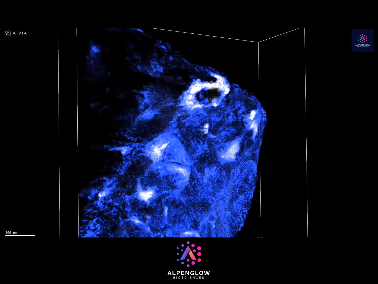
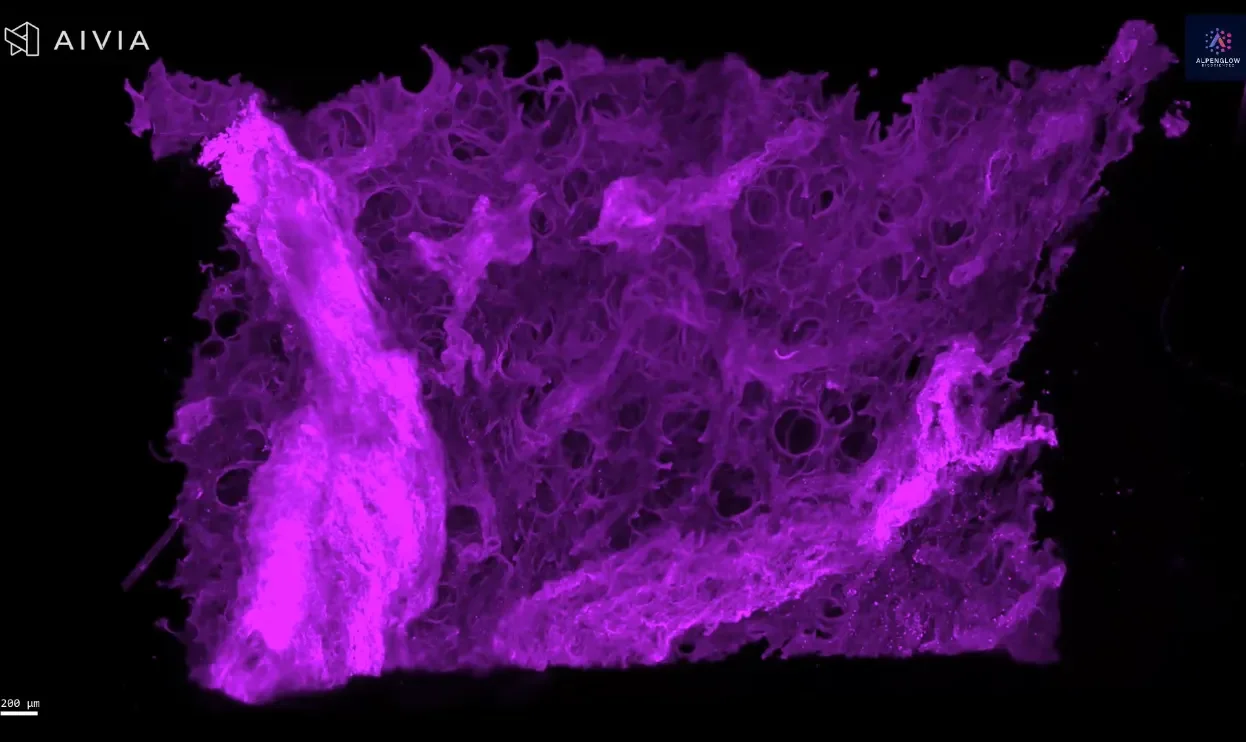
3D Imaging of Human Placenta Tissue Reveals PD-L1 at the Maternal Fetal Interface
This video presents a volumetric view of human placental tissue, with PD-L1 signal shown in cyan across the imaged tissue architecture.
PD-L1 is an immune-regulatory protein expressed in placental tissue and is relevant to research on trophoblast biology and the maternal-fetal interface. Viewing the signal in 3D preserves its distribution across depth and provides spatial context that cannot be captured from a single tissue section.
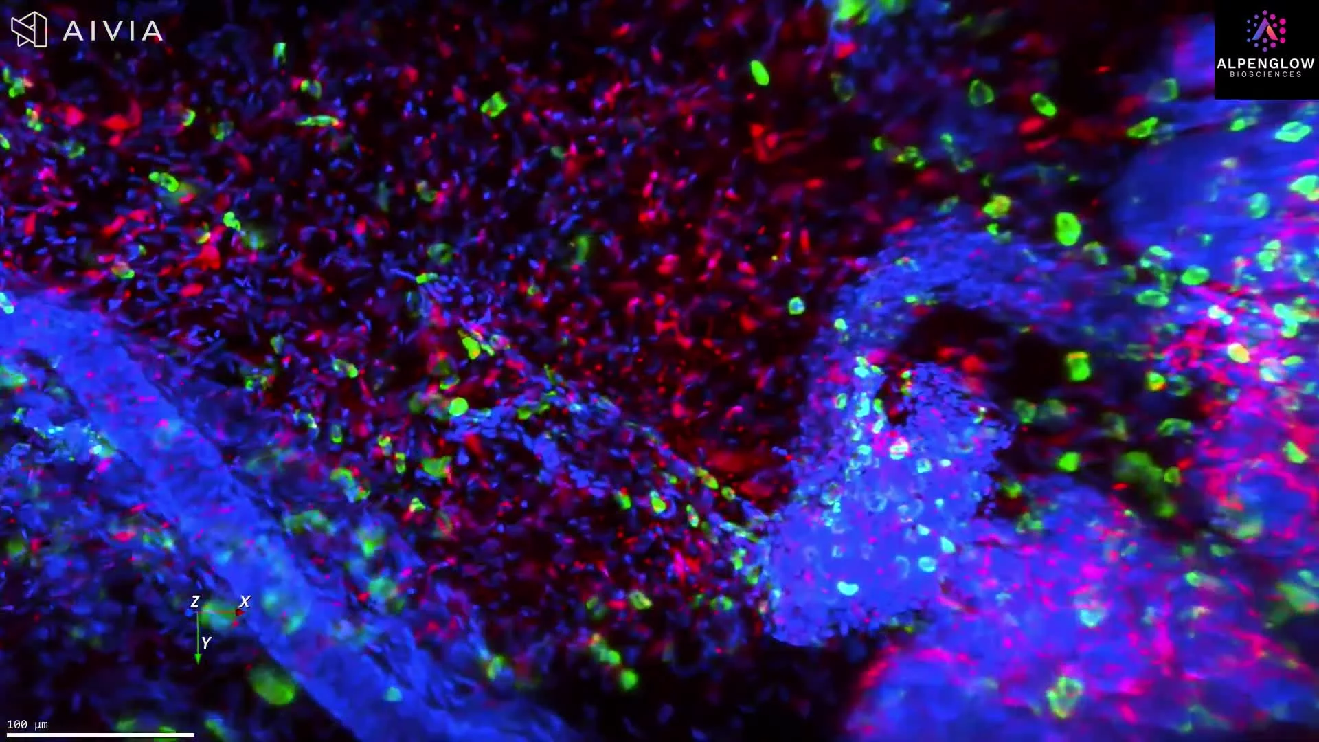
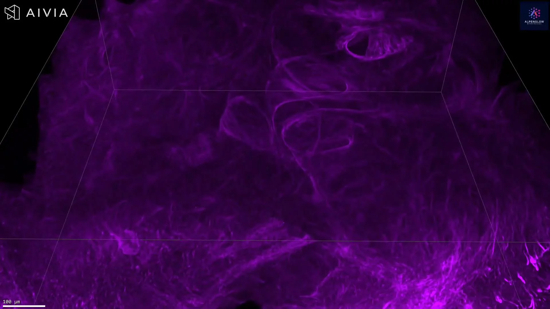
The volumetric dataset can support analysis of PD-L1-positive regions, signal distribution, tissue coverage, and spatial relationships with additional cellular or structural markers when included in a multiplex panel.
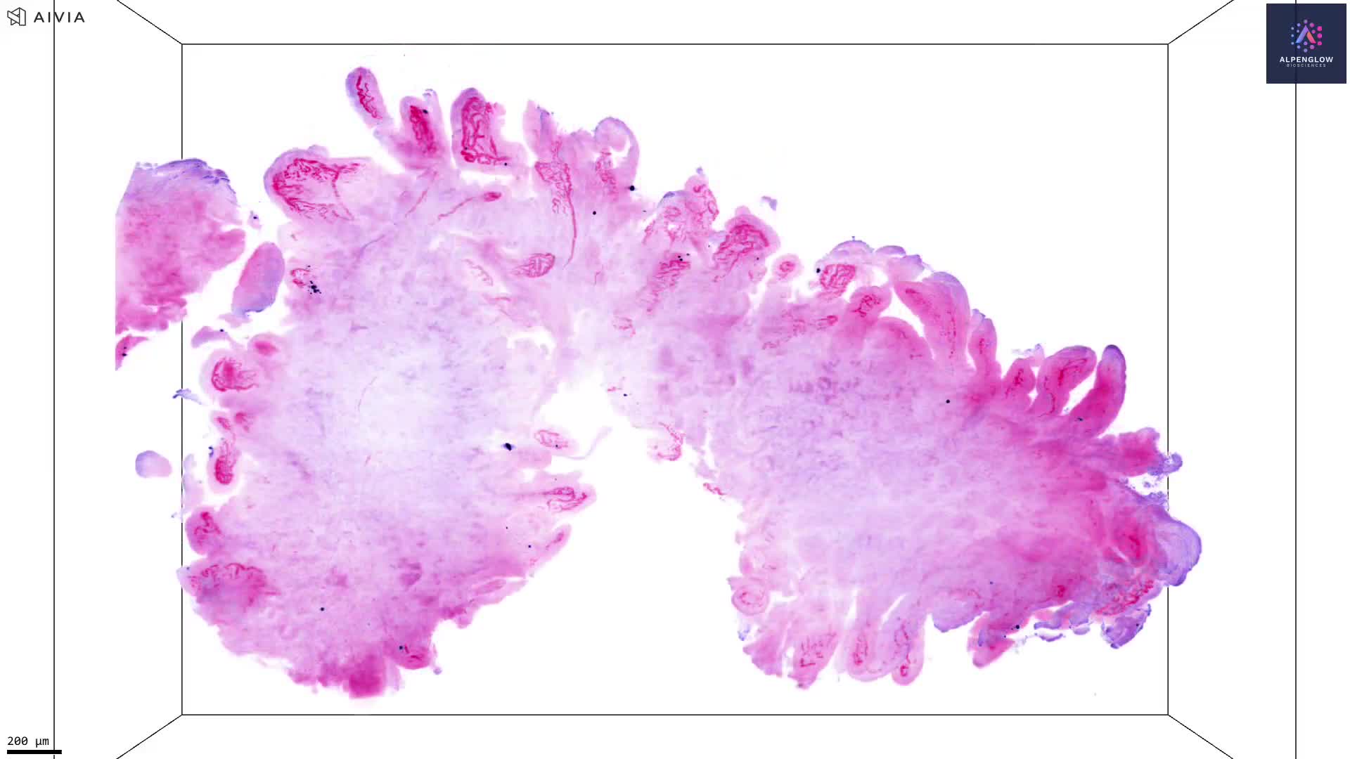
The tissue was imaged using the Aurora 3D™ Spatial Biology Solution, including the 3Di™ Hybrid Open-Top Light-Sheet microscope.