Summit AI™
Turn full-sample tissue imaging into measurable disease biology
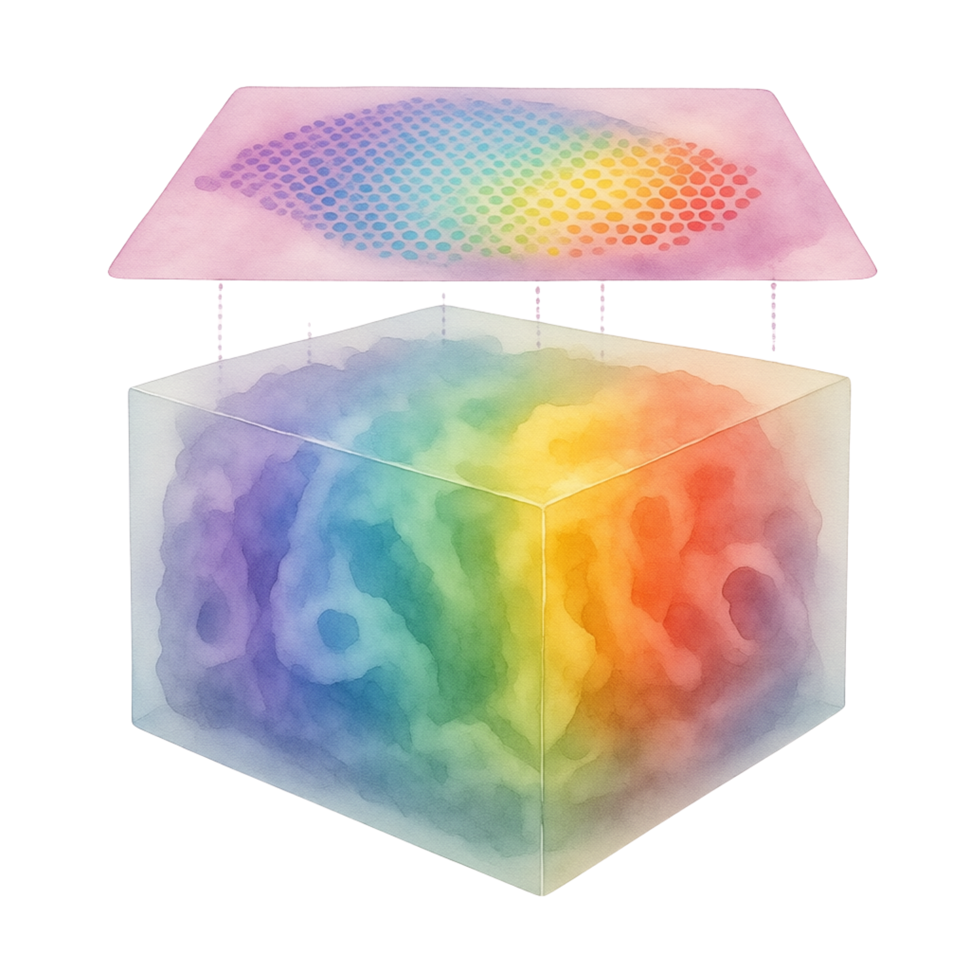
Summit AI™ analyzes full-sample imaging data and integrates multimodal data layers to generate quantitative tissue signatures that reflect how disease is organized across tissue.
Platform highlights
From full-sample imaging to quantitative tissue intelligence
Summit AI™ is designed to help teams move from large tissue images and volumetric datasets to measurable, confidence-aware outputs that support spatial profiling, digital pathology workflows, and quantitative tissue analysis.
Full-sample analysis
Measure tissue as a complete biological system, not as isolated regions or partial fields of view.
3D tissue signatures
Capture spatial organization across full tissue volume to support quantitative tissue analysis.
Uncertainty metrics
Interpret AI-powered analysis outputs with uncertainty metrics, confidence-aware review, and traceable image context.
Models that improve
Refine analysis through expert input and growing datasets to support repeatable workflows.
Why Summit AI™
From fragmented tissue analysis to full-sample quantitative insight
Current tissue analysis workflows often struggle with scale, iteration, disconnected tools, and inconsistent outputs. Summit AI™ brings these steps into a unified AI-powered analysis workflow for full-sample 3D tissue imaging and quantitative tissue analysis.
Analysis bottleneck
Tools do not scale
Large tissue images are difficult to analyze consistently across the full sample.
With Summit AI™
Scales to full-sample data
Analyze full-sample imaging data while preserving whole tissue context.
Analysis bottleneck
Iteration is slow
Reviewing outputs and changing parameters can slow analysis refinement.
With Summit AI™
Supports faster refinement
Expert input and repeatable workflows help teams refine outputs efficiently.
Analysis bottleneck
Workflows are fragmented
Review, segmentation, measurement, and interpretation happen across tools.
With Summit AI™
Unifies the pipeline
Connect image review, AI-powered analysis, spatial profiling, and measurement.
Analysis bottleneck
Outputs are hard to compare
Qualitative readouts can make cross-sample comparison difficult.
With Summit AI™
Focuses on insight
Generate measurable tissue signatures across samples and cohorts.
Multimodal context
Connect tissue images, data layers, and measurable disease biology
Summit AI™ is designed to connect full-sample 3D tissue imaging with complementary pathology, spatial biology, molecular, clinical, and study metadata, helping teams analyze tissue structure, spatial organization, and biological context at the sample and cohort level.
- 3D tissue imaging: volumetric tissue structure, morphology, and spatial organization across the full sample.
- Pathology and 2D tissue readouts: H&E and other image-based tissue data layers that support interpretation.
- Spatial and molecular biology: spatial transcriptomics, molecular maps, and marker-based readouts linked to tissue context.
- Clinical and study metadata: patient group, treatment, timepoint, lesion status, response, outcome, and cohort information connected to downstream analysis.
The result is an integrated analysis layer for quantitative tissue signatures, cross-sample comparison, and response-linked biological insights.
How Summit AI™ works
From tissue data to quantitative spatial evidence
Summit AI™ connects full-sample 3D tissue imaging, complementary data layers, expert input, and AI-driven analysis to turn complex tissue context into structured measurements, maps, cohort comparisons, and source-linked outputs.
Built around confidence-linked analysis. The workflow keeps image context connected to segmentation, measurement, and interpretation so teams can trace outputs back to the source tissue data.
Data ingestion
ConnectIngest 3D tissue volumes together with relevant pathology, spatial biology, molecular, clinical, and study metadata.
Target definition and expert input
DefineDefine biological targets, tissue regions, cell types, structures, and study questions with expert guidance. Annotations and corrections can refine downstream analysis.
Biological object segmentation
SegmentSegment cells, vessels, nerves, follicles, remodeled regions, and complex tissue boundaries across the 3D tissue volume.
Spatial feature extraction
ExtractConvert segmented objects and regions into quantitative features, including morphology, intensity, volume, surface area, branching, density, distances, neighborhoods, and cell-to-structure relationships.
Agentic analysis and outputs
QueryTranslate biological questions into structured analysis workflows that return measurements, visual overlays, cohort comparisons, and source-linked reports.
AI-guided analysis
From biological questions to structured spatial analysis
Summit AI™ uses an agentic analysis layer to translate biological questions into structured workflows across segmented 3D tissue objects, extracted spatial features, cohort metadata, and source-linked image context.
Spatial Statistics Agent
Ask tissue-level questions in biological language
Query cells, structures, regions, spatial relationships, and cohort-level patterns without separating the analysis from the original tissue data.
“Are immune cells enriched near nerves, follicles, vessels, or remodeled regions in treated versus untreated tissue?”
Output: segmented objects, spatial measurements, visual overlays, cohort comparisons, and source-linked regions for review.
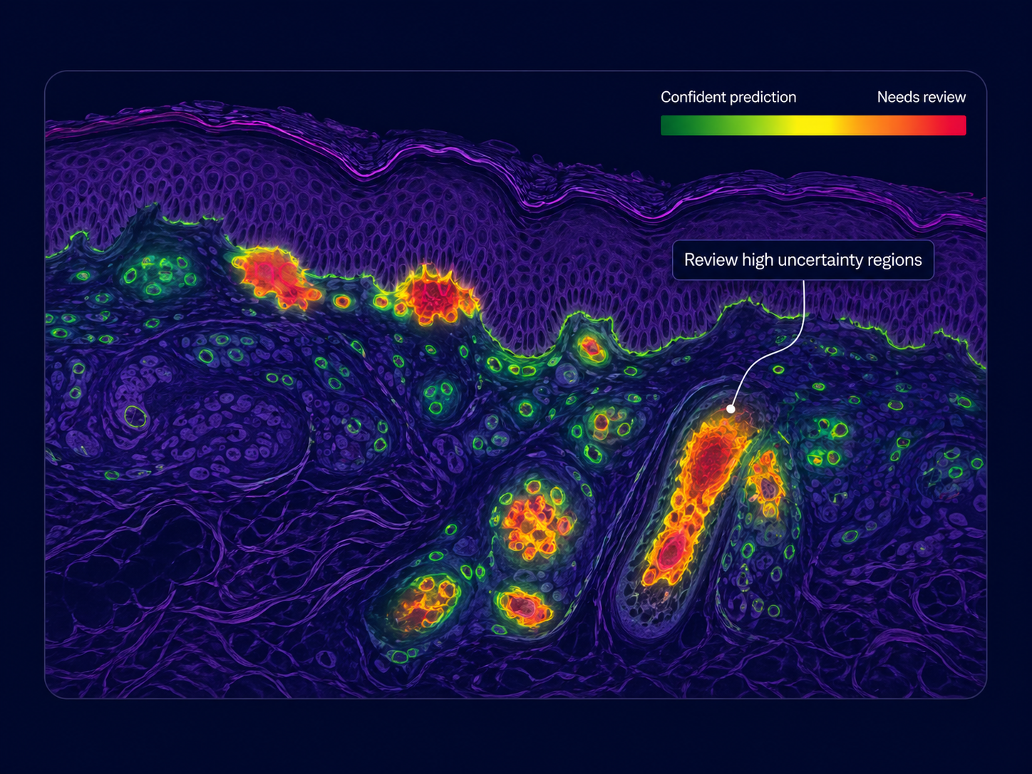
Know where review matters
Confidence-aware outputs help identify cells, structures, or regions that may require expert review, while keeping predictions, measurements, and edits linked back to the source image.
Quantitative outputs
Turn image data into structured tissue readouts
Volume level
3D tissue signatures
Spatial organization captured across full tissue volume and linked to quantitative tissue analysis.
Disease level
Disease signatures
Measurable tissue patterns that help characterize how disease is organized across samples.
Feature level
Feature-level measurements
Structured measurements across cells, structures, regions, and tissue features.
Cohort level
Cohort-level comparisons
Compare tissue signatures and spatial patterns across samples and groups.
Review level
Confidence-aware outputs
Uncertainty metrics help highlight outputs and regions that may need expert review.
Trace level
Image-traceable results
Keep quantitative readouts connected to the source image and tissue context.
Initial deployment area
Built first for inflammatory skin disease research
Summit AI™ is first deployed in dermatology, where full biopsy context can help teams evaluate tissue architecture, immune organization, barrier disruption, and structural remodeling across inflammatory skin disease research.
Why full-sample analysis matters: skin biology is spatially organized across tissue layers, epithelial structures, immune neighborhoods, nerves, follicles, and remodeling patterns.
Dermatology readouts
Full biopsy context
Analyze intact skin biopsy volumes with whole tissue context.
Quantitative tissue features
Measure architecture, immune patterns, and remodeling in 3D tissue imaging datasets.
Extensible framework
Apply the same AI-powered analysis workflow to additional tissue-driven indications.
Models grow with data
A learning layer that becomes more useful as evidence accumulates
Summit AI™ connects expert review, image-linked feedback, and reusable structured outputs so analysis can be refined as 3D tissue imaging datasets grow.
From review to refinement to reuse.
Review
Inspect outputs in the original tissue context.
Feedback
Capture confirmations, edits, and expert input.
Refine
Improve repeatable segmentation and measurement.
Reuse
Apply structured readouts across samples and studies.
See Summit AI™ in action
Turn tissue context into measurable disease biology
Explore how Summit AI™ connects full-sample 3D tissue imaging, AI-powered analysis, spatial profiling, and digital pathology workflows.
A connected workflow for tissue intelligence
- ✓ Analyze full-sample 3D tissue imaging data.
- ✓ Generate quantitative tissue signatures.
- ✓ Review outputs with image-linked context.