
Dermatology Investigator
3D spatial biology for dermatology research
Measure skin biology across the full tissue volume
Visualize intact skin architecture, peripheral nerves, immune cells, vessels, follicles, and neuroimmune relationships in 3D. Alpenglow combines whole-tissue imaging with AI-powered analysis to generate quantitative spatial readouts for inflammatory skin disease, hair disorders, oncology, wound healing, and treatment-response research.
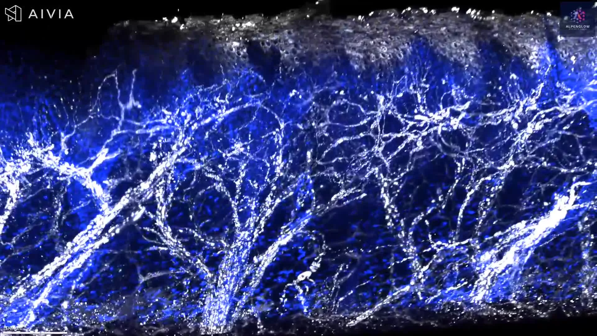
Volumetric 3D imaging of rat toe pad skin reveals cutaneous nerve fibers, branching, and terminal structures at the dermal–epidermal junction using PGP9.5 and YO-PRO-1 staining.
This dataset presents volumetric 3D imaging of rat toe pad skin, revealing cutaneous nerve architecture at the dermal–epidermal junction.
PGP9.5 highlights nerve fibers in white, while YO-PRO-1 labels nuclei in blue. The intact tissue volume preserves the continuity of larger nerve bundles, finer branches, fiber crossings, and terminal structures across tissue depth.
With appropriate segmentation, the dataset can support quantitative analysis of nerve density, fiber length, branching, orientation, and the distribution of nerve fibers relative to the epidermis and surrounding cells. These measurements are relevant to studies of cutaneous innervation, sensory biology, neuro-immune interactions, pain, itch, and disease-associated changes in nerve architecture.
The tissue was imaged using the Aurora 3D™ Spatial Biology Solution, including the 3Di™ Hybrid Open-Top Light-Sheet microscope.
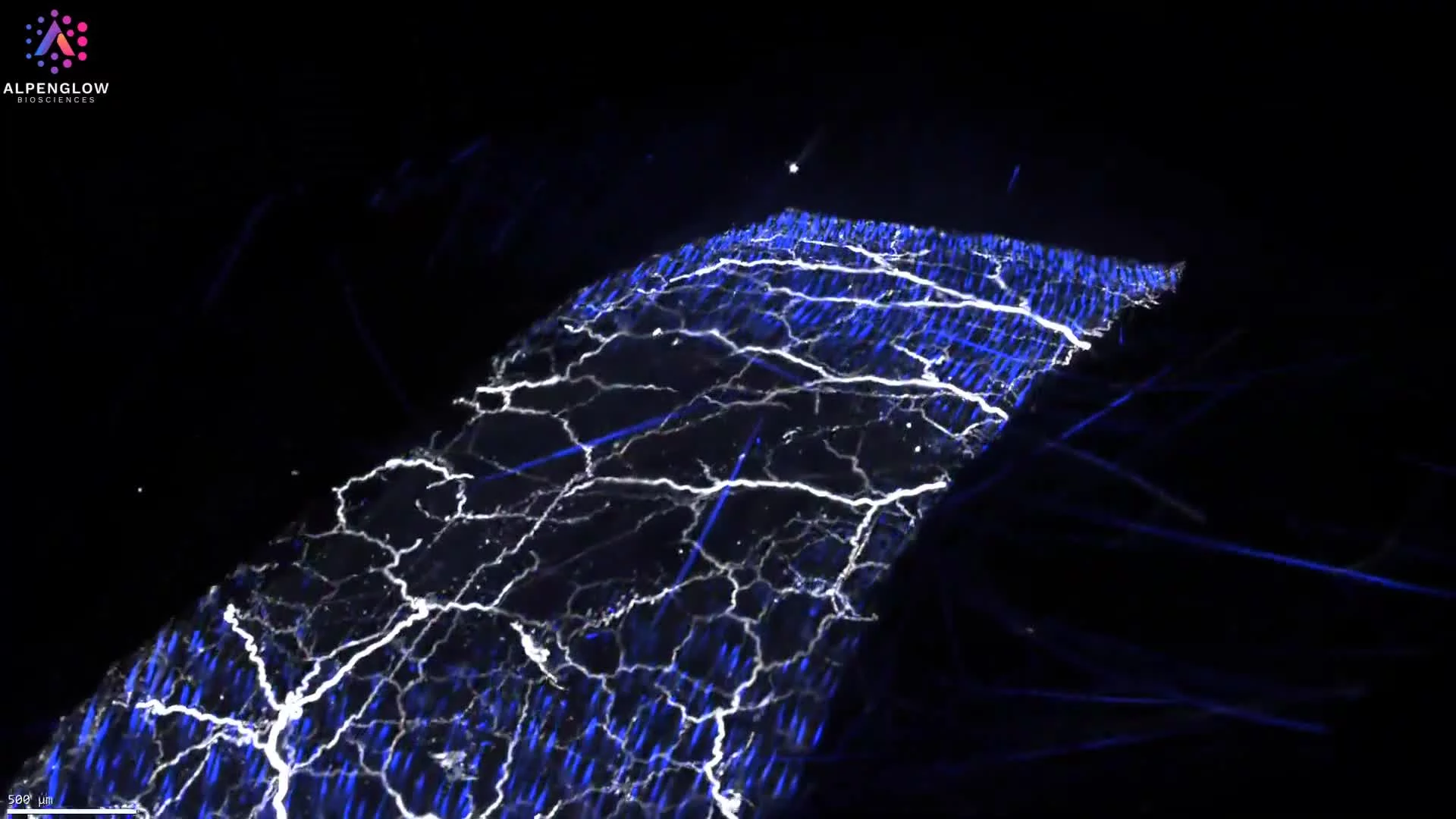
3D imaging of mouse skin reveals how larger dermal nerve bundles divide into progressively finer branches across the tissue volume. YO-PRO-1 labels nuclei, while PGP9.5 highlights cutaneous nerve architecture.
This dataset presents mouse skin tissue imaged in 3D, with YO-PRO-1 labeling nuclei and PGP9.5 highlighting cutaneous nerve structures.
The volumetric view reveals larger nerve bundles within the dermis as they divide into progressively finer branches and projections across the imaged tissue volume. Preserving these structures in 3D makes it possible to follow nerve continuity, branching patterns, orientation, and relationships with surrounding cells and tissue compartments.
With appropriate segmentation, the dataset can support measurement of nerve density, fiber length, branching frequency, tortuosity, orientation, and regional variation. These features are relevant to research on cutaneous innervation, sensory biology, neuro-immune interactions, pain, itch, and changes associated with disease or treatment.
The tissue was imaged using the Aurora 3D™ Spatial Biology Solution, including the 3Di™ Hybrid Open-Top Light-Sheet microscope.
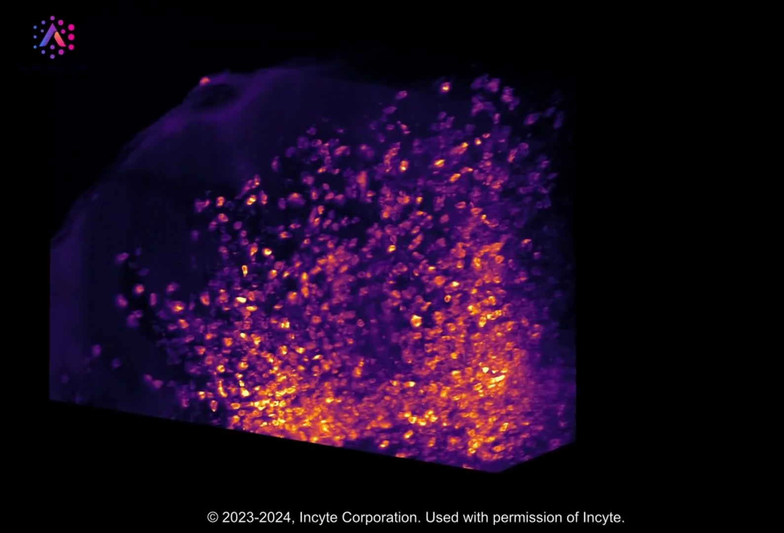
High-resolution 3D imaging of CD45-stained skin biopsy shows lymphocyte clustering around nerves, offering new insights into immune cell behavior.
This dataset showcases a whole skin punch biopsy stained for CD45, imaged with cutting-edge 3D technology.
The visualization highlights the spatial distribution of lymphocytes within the epidermis and dermis, revealing how immune cells cluster around nerve networks and integrate into the skin’s complex architecture.
Advanced data managementand AI-powered segmentation enable precise mapping of lymphocyte distribution, capturing patterns invisible to conventional 2D histology.
These results not only provide stunning imagery but also uncover fundamental insights into immune–neural interactions in skin, fueling progress in dermatology research and reshaping our understanding of inflammatory skin disease.
By combining whole-tissue 3D imaging with quantitative analysis, this work demonstrates the transformative potential of digital pathology and spatial biology.
See our AD use case.
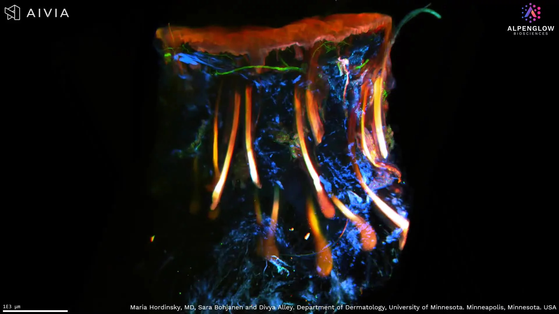
High-resolution 3D imaging of scalp tissue reveals nuclei, immune cells, and nerve networks, driving new insights into alopecia and other skin diseases.
This high-resolution 3D visualization of scalp tissue provides unprecedented clarity into structures relevant to hair biology and disease. The dataset captures the epidermal layer, peripheral nerve distribution, and piliferous bulb architecture in measurable detail, features critical to understanding alopecia and related conditions.
Stains used:
TO-PRO-3 (Red): Highlights nuclei in the epidermis
CD45 (Blue): Marks immune cell distribution
PGP9.5 (Green): Maps peripheral nerve innervation
By comparing volumetric 3D analysis with conventional 2D slices, this dataset demonstrates the significant loss of information in single-plane histology, underscoring the value of whole-tissue imaging with HOTLS microscopy. Leveraging advanced data management and AI-powered segmentation, immune and neural structures are quantified with precision.
The insights extend beyond visualization, contributing to dermatology research in alopecia areata, prurigo nodularis, hidradenitis suppurativa, atopic dermatitis (see our AD use case), and other inflammatory skin diseases.
These breakthroughs highlight the transformative potential of 3D spatial biology and digital pathology in uncovering the mechanisms underlying hair loss and skin disorders.
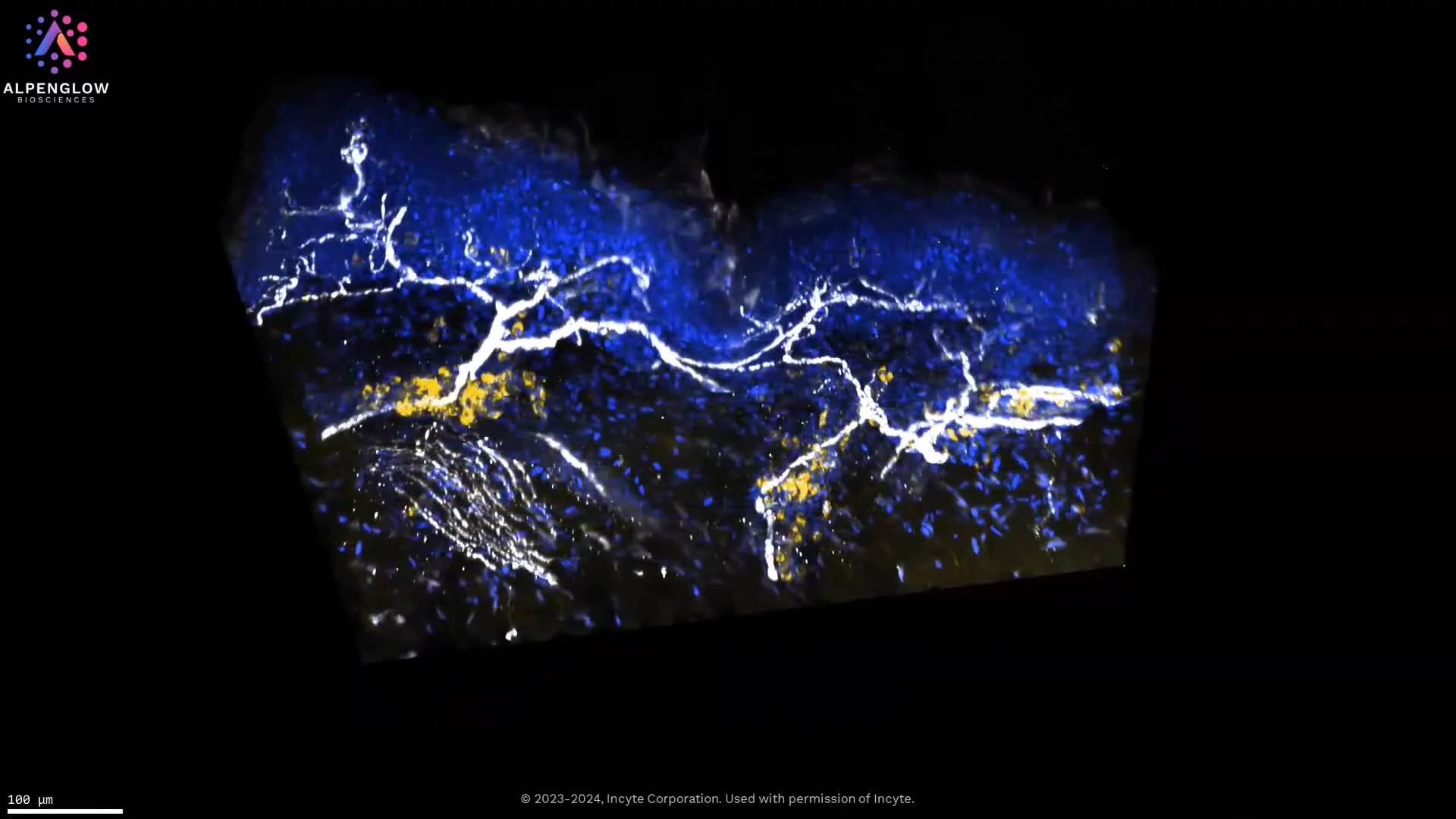
High-resolution 3D imaging of atopic dermatitis reveals lymphocyte clusters near nerves, stained with TO-PRO-3, PGP 9.5, and CD45. Explore detailed innervation and immune-cell interactions in lesional skin.
This high-resolution 3D visualization of lesional atopic dermatitis skin tissue reveals the spatial relationship between cutaneous nerves and CD45-positive immune cells.
Using high-resolution 40X HOTLS imaging, TO-PRO-3, shown in blue, labels nuclei. PGP9.5, shown in white, traces nerve fibers, while CD45, shown in yellow, highlights immune cells across the tissue volume.
The volumetric view preserves epidermal and dermal architecture while revealing clusters of CD45-positive immune cells near selected nerve fibers across tissue depth. This spatial proximity provides a tissue-wide view of neuro-immune organization without implying a functional interaction from imaging alone.
With appropriate segmentation, the dataset supports measurement of immune-cell density, nerve density, clustering, distances between immune cells and nerves, and regional variation across skin compartments. These readouts are relevant to research on inflammatory skin diseases and the neuroimmune mechanisms underlying itch and inflammation.
Explore how 3D tissue imaging supports dermatology research across skin architecture, innervation, immune organization, and spatial relationships.
The tissue was imaged using the Aurora 3D™ Spatial Biology Solution, including the 3Di™ Hybrid Open-Top Light-Sheet microscope.
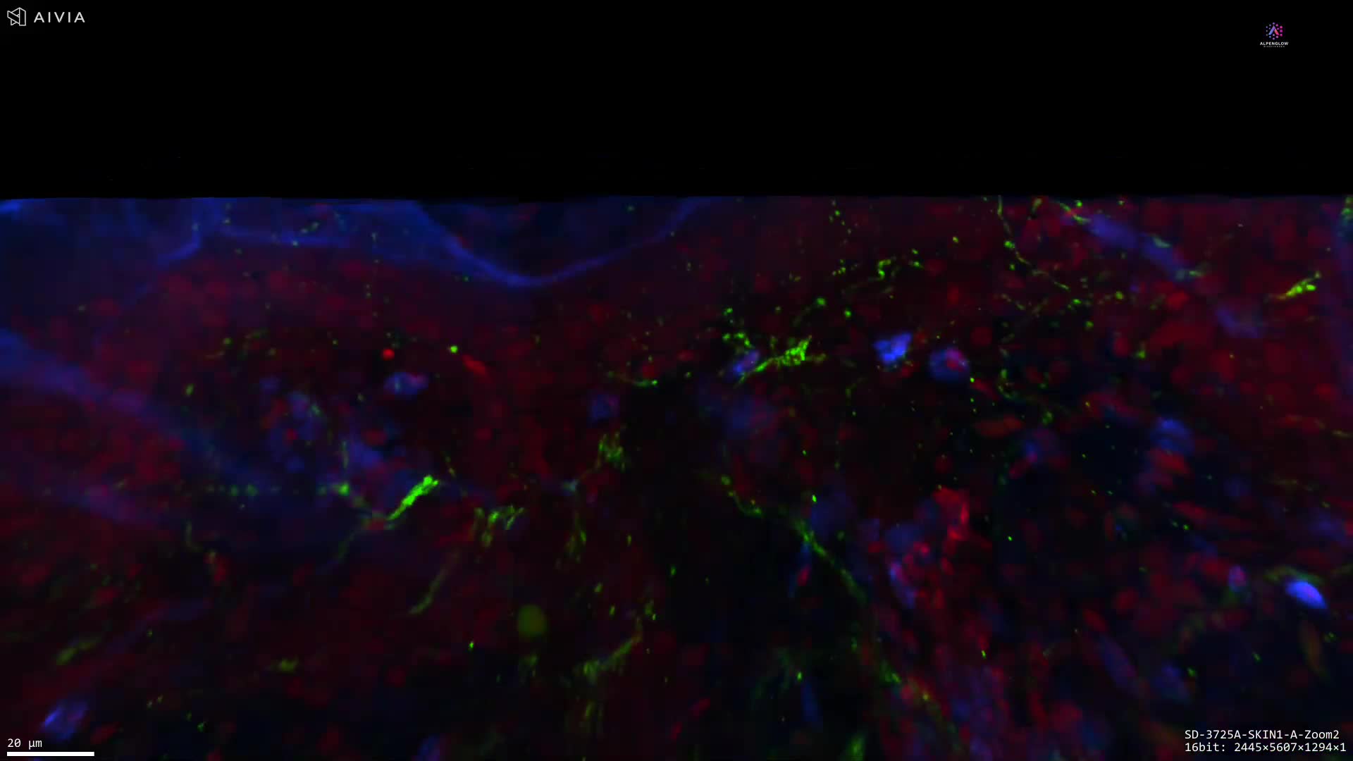
High-resolution 3D imaging of human skin preserves hair follicle, glandular, and neural architecture across depth, supporting spatial and quantitative analysis for hair and dermatology research.
This high-resolution 3D dataset presents human skin tissue, preserving the architecture of a hair follicle, its associated gland, and the surrounding nerve network across depth.
The volumetric view allows the follicle and glandular structure to be followed through the tissue, revealing their morphology, orientation, and spatial relationships with nearby nerve fibers. This continuous view reduces the fragmentation of complex structures that can occur across individual 2D tissue sections.
Following processing with 3Dm™ data management and analysis using 3Dai™ AI-powered analysis, the dataset supports measurement of follicle dimensions, glandular morphology, nerve density, branching, and distances between neural and follicular structures. These readouts are relevant to research on alopecia, inflammatory skin conditions, hair biology, and treatment-associated changes in follicular architecture.
Explore how 3D tissue imaging supports dermatology research across skin architecture, innervation, immune organization, and spatial relationships.
The tissue was imaged using the Aurora 3D™ Spatial Biology Solution, including the 3Di™ Hybrid Open-Top Light-Sheet microscope.
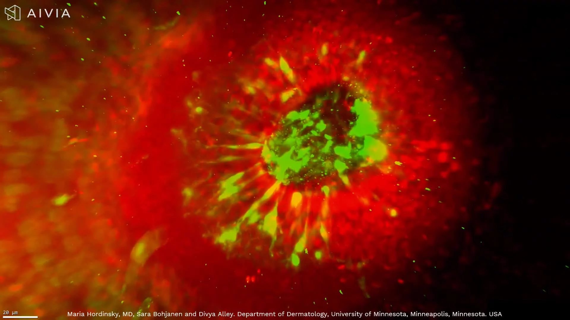
See hair follicles in true 3D, from bulb to dermal papilla, with detailed mapping of sensory nerves and nuclei for alopecia and hair research.High-resolution 3D imaging of scalp tissue reveals nuclei, immune cells, and nerve networks, driving new insights into alopecia and other skin diseases.
For the first time, a hair follicle can be visualized in its true three-dimensional architecture, from the bulb where growth begins to the dermal papilla, a key signaling center for follicle development. This high-resolution dataset, captured with the Aurora platform and HOTLS microscopy, also reveals the hair plexus, a dense network of sensory nerve fibers critical for touch sensation.
Stains used:
PGP9.5 (Green): Sensory nerve fibers surrounding the follicle
TO-PRO-3 (Red): Cell nuclei distribution
Through advanced 3D fluorescence imaging, combined with data management and AI-powered segmentation, subtle structural changes in the follicle can be detected earlier than with conventional 2D histology. This capability is transforming alopecia research, providing a new window into hair biology, disease mechanisms, and treatment response.
By integrating quantifiable 3D tissue imaging with digital pathology and spatial profiling, researchers gain unprecedented insights into hair follicle biology and inflammatory skin conditions. This breakthrough demonstrates how better imaging leads directly to better insights, and ultimately better treatments.
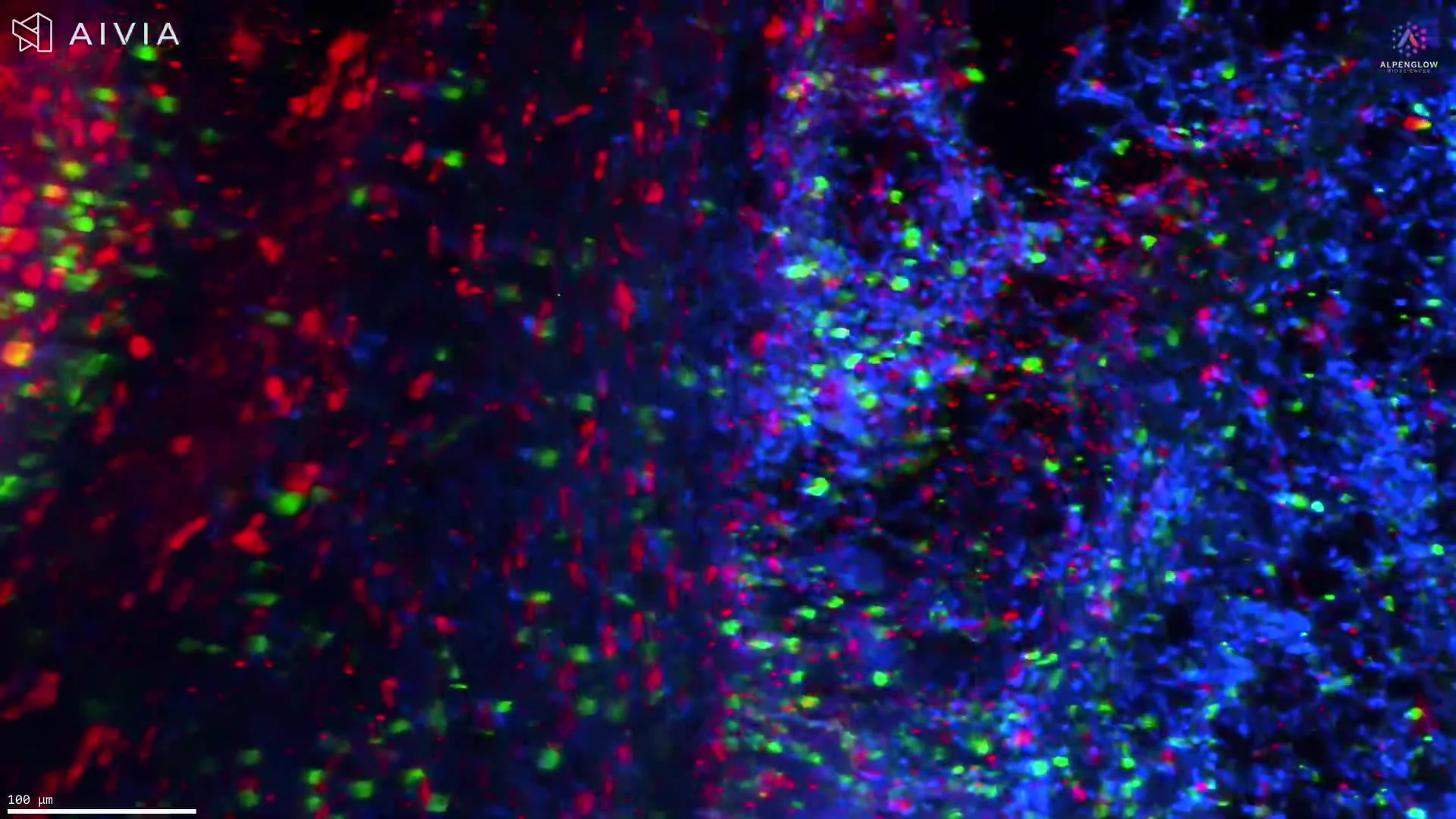
3D imaging of melanoma tissue with CD45, Neutrophil Elastase, and TO-PRO-3 reveals hidden immune–tumor interactions and sets a new standard for research.
Through advanced staining and high-resolution 3D imaging, we move beyond the limitations of flat, 2D slides to capture the full spatial context of human melanoma tissue.
In this video, you can observe:
🟢 CD45 – highlighting immune cells
🔴 Neutrophil Elastase – marking neutrophils
🔵 TO-PRO-3 – illuminating nuclei
Each marker reveals cellular architecture with striking clarity, uncovering complex immune–tumor interactions previously hidden in 2D views.
This depth of visualization enhances our understanding of melanoma biology and establishes a new benchmark for research.
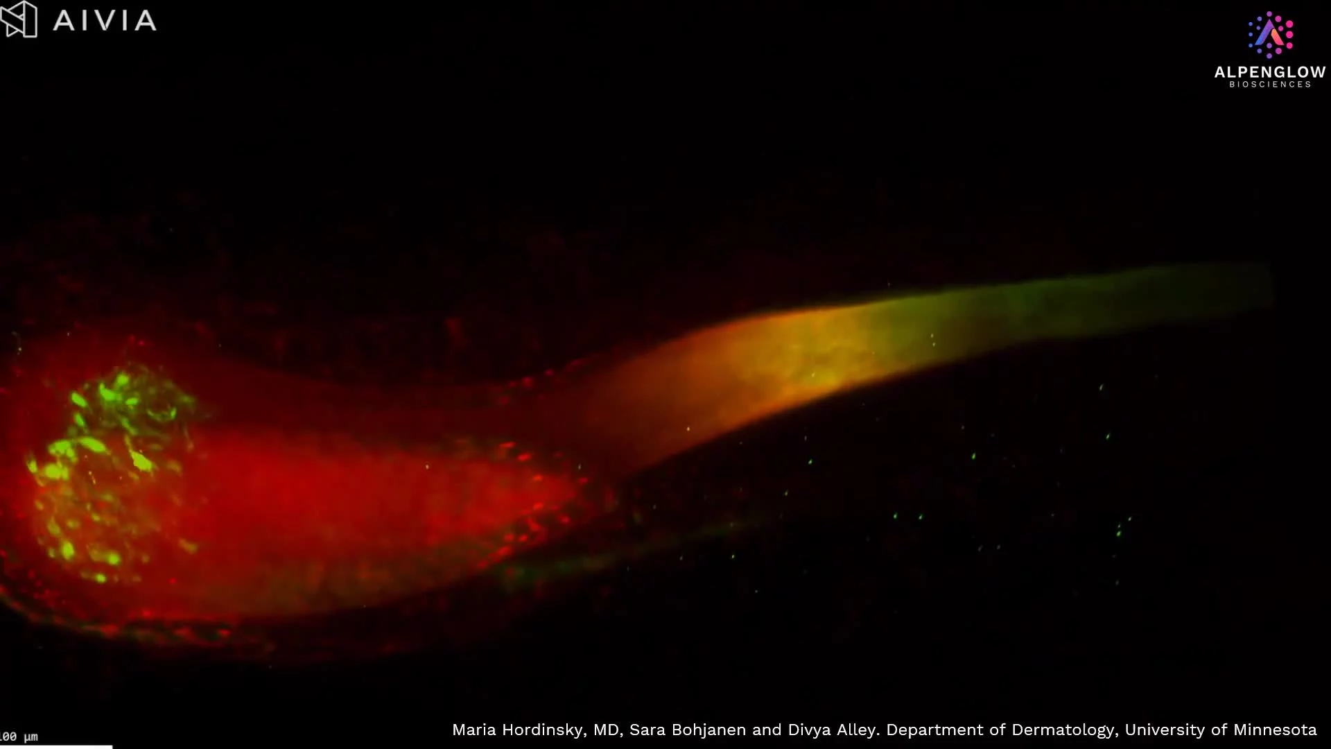
See an entire hair follicles in true 3D, from bulb to dermal papilla, with detailed mapping of sensory nerves and nuclei for alopecia and hair research.High-resolution 3D imaging of scalp tissue reveals nuclei, immune cells, and nerve networks, driving new insights into alopecia and other skin diseases.
For the first time, a hair follicle can be visualized in its true three-dimensional architecture, from the bulb where growth begins to the dermal papilla, a key signaling center for follicle development. This high-resolution dataset, captured with the Aurora platform and HOTLS microscopy, also reveals the hair plexus, a dense network of sensory nerve fibers critical for touch sensation.
Stains used:
PGP9.5 (Green): Sensory nerve fibers surrounding the follicle
TO-PRO-3 (Red): Cell nuclei distribution
Through advanced 3D fluorescence imaging, combined with data management and AI-powered segmentation, subtle structural changes in the follicle can be detected earlier than with conventional 2D histology. This capability is transforming alopecia research, providing a new window into hair biology, disease mechanisms, and treatment response.
By integrating quantifiable 3D tissue imaging with digital pathology and spatial profiling, researchers gain unprecedented insights into hair follicle biology and inflammatory skin conditions. This breakthrough demonstrates how better imaging leads directly to better insights, and ultimately better treatments.
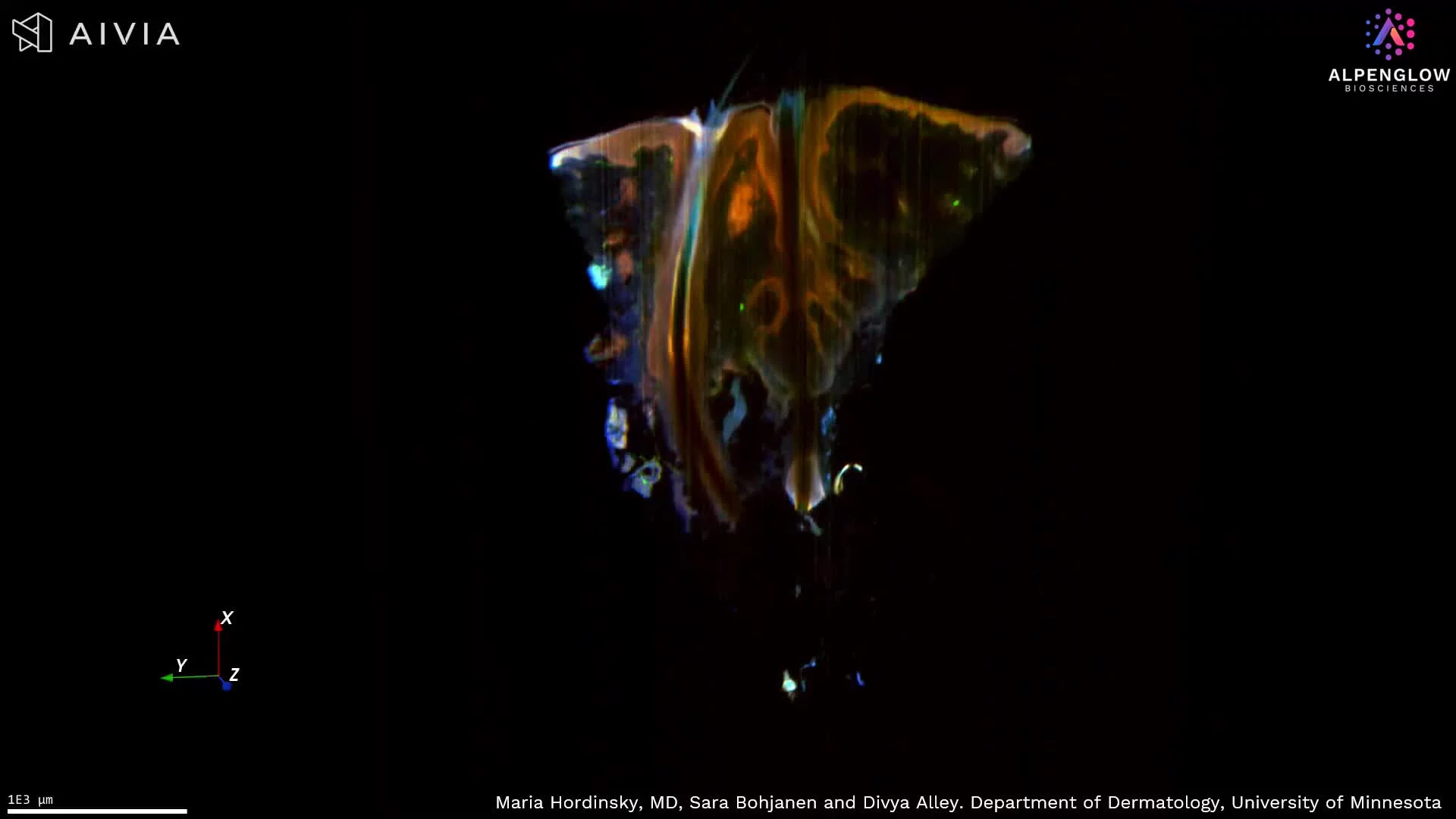
High-resolution 3D imaging of scalp tissue preserves hair follicle architecture, immune-cell distribution, and cutaneous nerve networks across depth, supporting spatial and quantitative analysis for hair and inflammatory skin research.
This high-resolution 3D dataset reveals scalp tissue architecture relevant to hair biology and inflammatory skin disease.
TO-PRO-3, shown in red, labels nuclei and highlights the epidermal and follicular cellular architecture. CD45, shown in blue, reveals the distribution of immune cells, while PGP9.5, shown in green, maps cutaneous nerve fibers across the tissue volume.
The volumetric view preserves intact hair follicles, including the hair bulb, along with surrounding immune cells and nerve networks. Following these structures through depth provides spatial information on follicle morphology, innervation, immune cell distribution, and relationships among skin compartments that may be fragmented in individual tissue sections.
The dataset supports quantitative analysis of follicle dimensions and orientation, immune-cell density and clustering, nerve density and branching, and distances between follicles, immune cells, and nerve fibers. These measurements are relevant to research on alopecia, atopic dermatitis, prurigo nodularis, hidradenitis suppurativa, and other inflammatory skin conditions.
Explore how 3D tissue imaging supports dermatology research across skin architecture, innervation, immune organization, and spatial relationships.
The tissue was imaged using the Aurora 3D™ Spatial Biology Solution, including the 3Di™ Hybrid Open-Top Light-Sheet microscope.
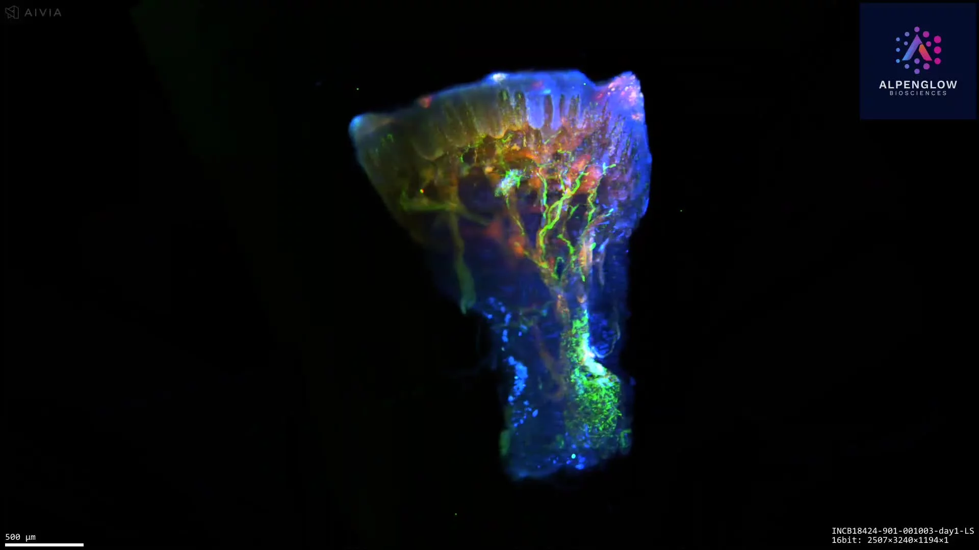
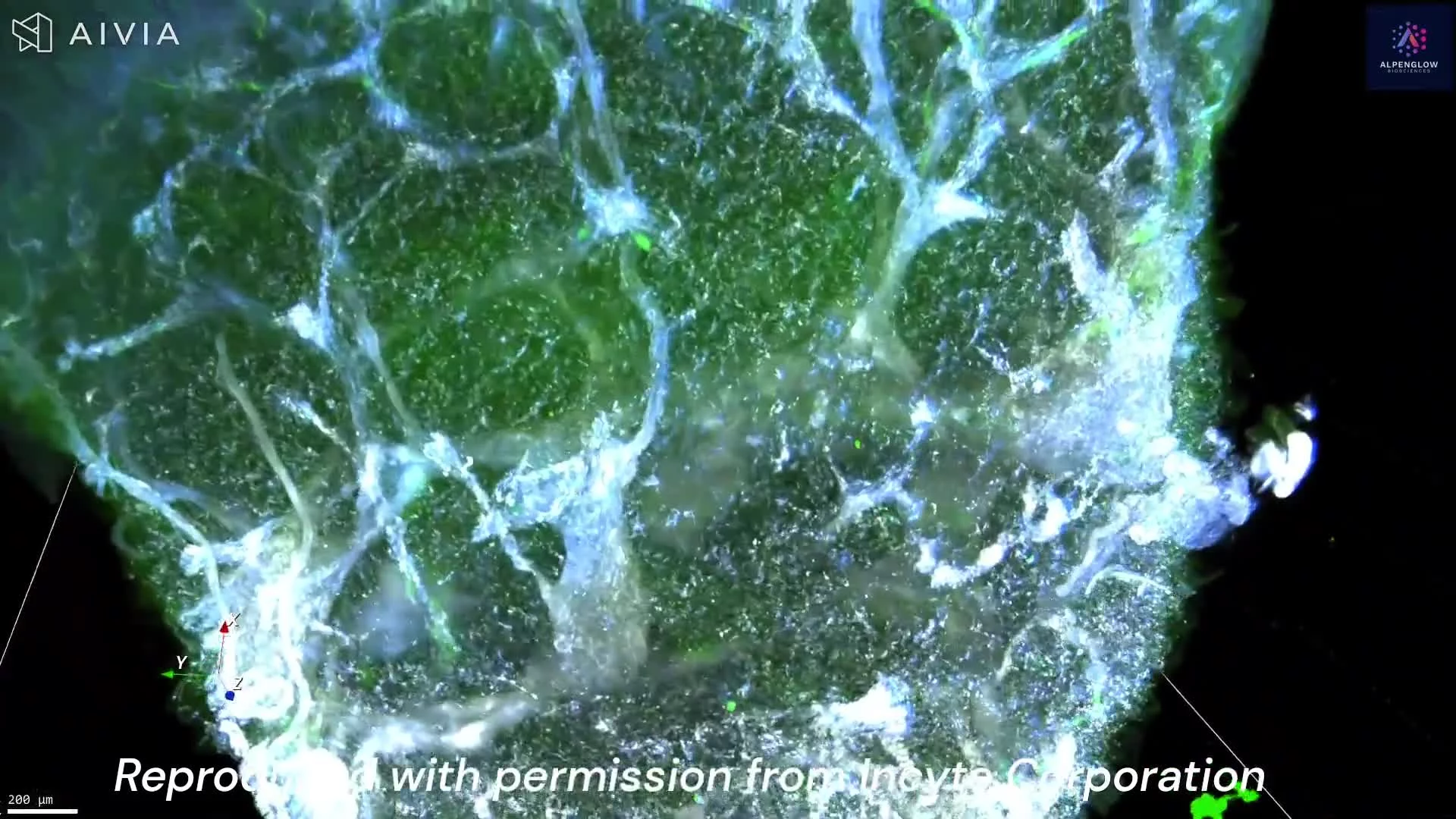
3D imaging of atopic dermatitis skin punch biopsy; the tissue is stained with TO-PRO-3, PGP9.5, and CD45. Explore detailed innervation and immune-cell interactions in lesional skin.
Low-resolution 3D imaging with Hybrid Open Top Light Sheet (HOTLS) microscopy of an entire lesional Atopic Dermatitis skin punch biopsy highlights epidermal and dermal innervation.
The tissue was stained with TO-PRO-3 (red, nuclei), PGP9.5 (green, nerves), and CD45 (blue, T cells), enabling comprehensive visualization of neuro-immune interactions and inflammatory changes across the full biopsy.
See our AD use case.
Image reproduced with permission from Incyte Corporation.
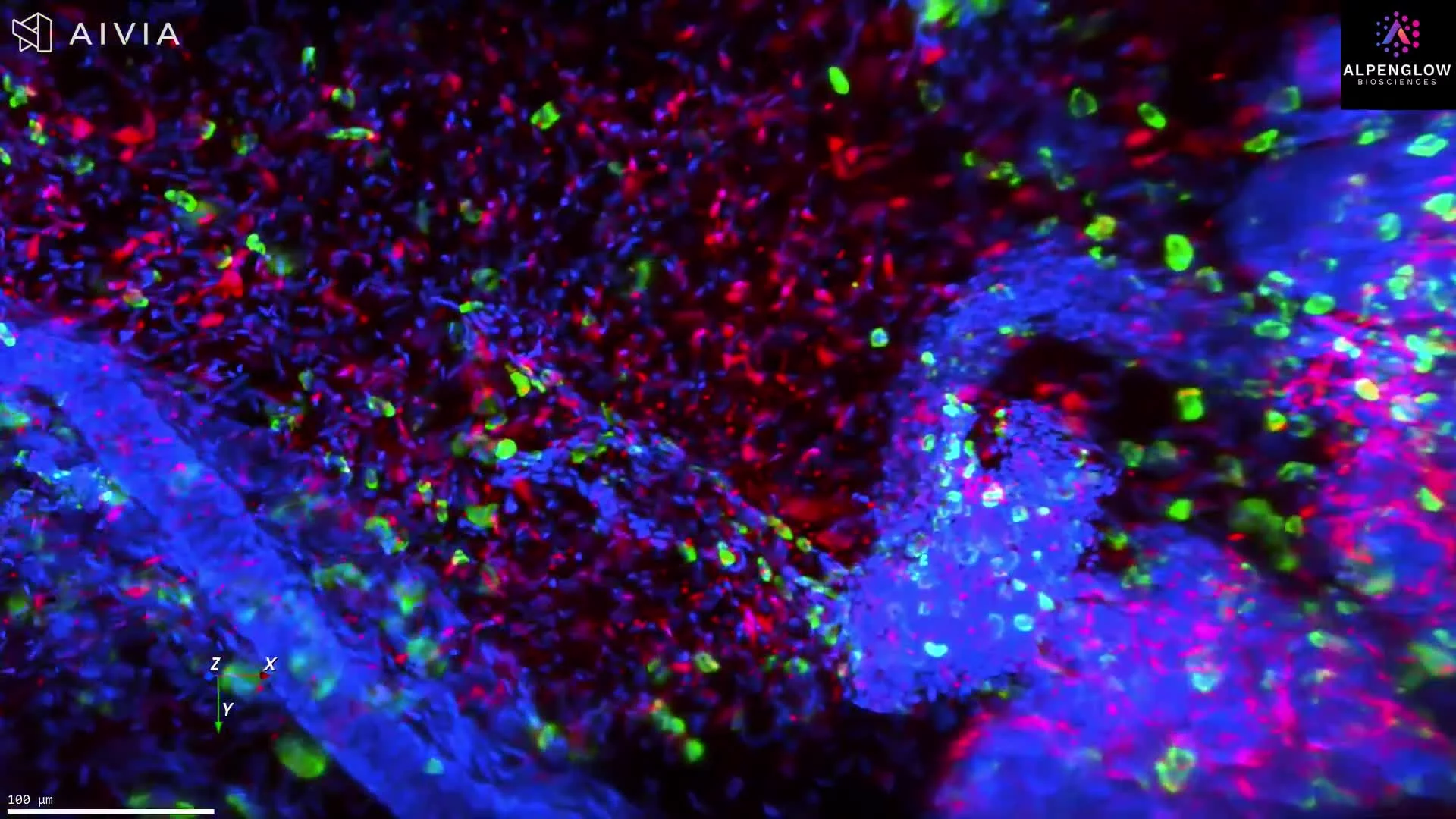
3D imaging of human skin biopsy stained with tryptase, TO-PRO-3, and PGP9.5 reveals mast cell–nerve interactions for dermatology and oncology research.
This dataset presents an intact, fluorescence-labeled human skin biopsy imaged in true 3D with the Aurora™ 3Di Hybrid Open Top Light Sheet (HOTLS) microscope. The volumetric view reveals the native architecture of neuroimmune interactions that conventional 2D slices cannot capture.
Stains used:
Tryptase (Green): Labels mast cells
TO-PRO-3 (Blue): Marks nuclei
PGP9.5 (Red): Traces nerves
By preserving full cell morphology and spatial relationships, this dataset provides the ground truth for quantifying mast cell density, mapping nerve proximity, and investigating mechanisms underlying chronic itch, fibrosis, and inflammatory skin disorders.
With integration of 3Dm data management and 3Dai AI-powered segmentation, researchers can perform reproducible, high-content quantification of neuroimmune interactions. This 3D approach eliminates slice bias and ensures accurate analysis at scale.
Applications span translational dermatology, where mast cells play a role in inflammatory skin disease, and immuno-oncology, where mast cell–nerve dynamics may influence tumor microenvironments. This example illustrates how 3D histology and digital pathology offer actionable insights that extend beyond visualization to measurable data.
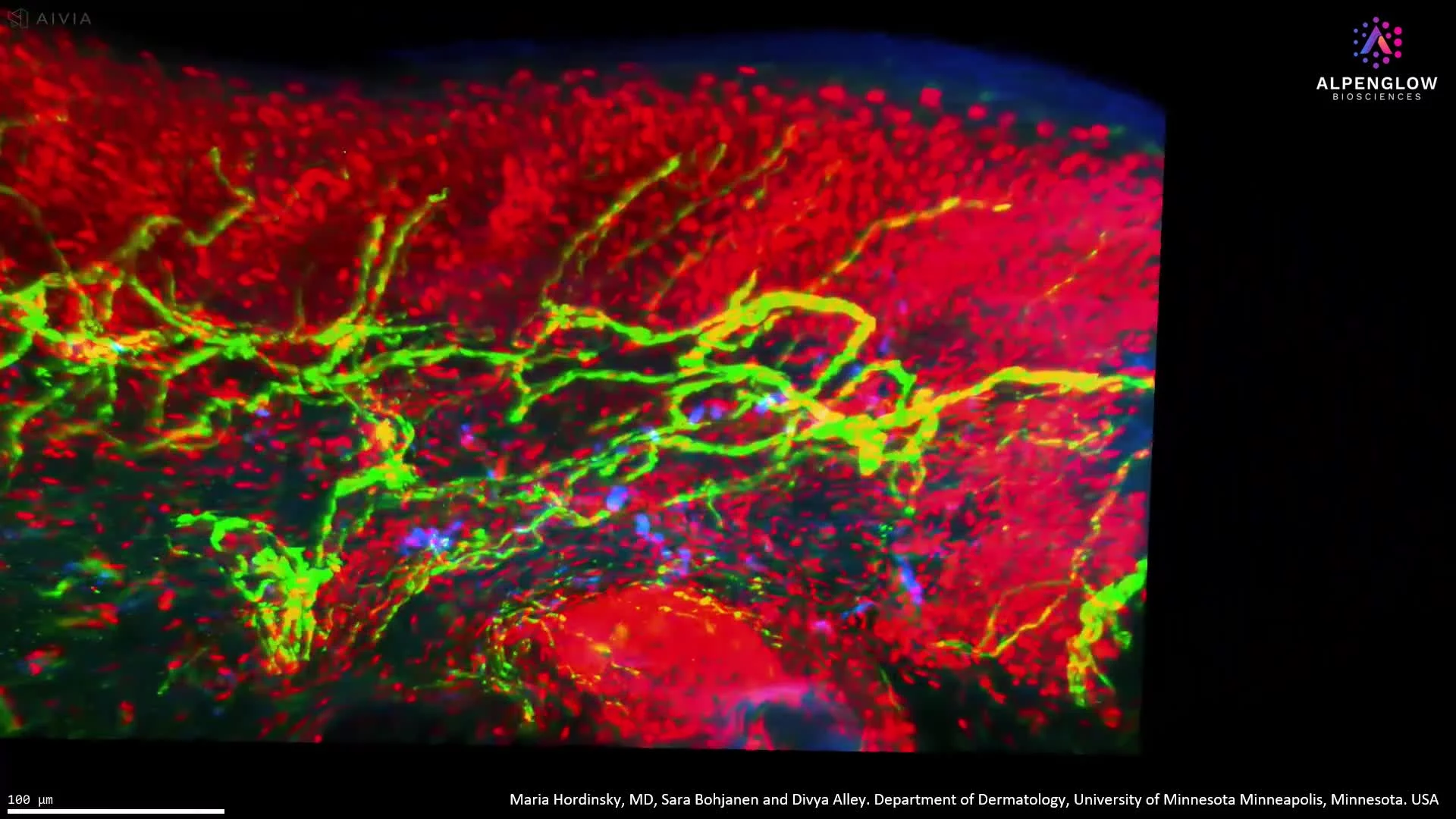
High-resolution 3D imaging of scalp epidermis reveals branching nerve structures and nuclear organization, advancing hair and dermatology research.
This stunning 3D visualization of the human scalp highlights the intricate patterns of epidermal innervation. Using HOTLS microscopy on the Aurora platform, the dataset captures how large peripheral nerves branch upward into fine structures that extend through the upper epidermis, creating a detailed map of nerve distribution.
Stains used:
TO-PRO-3 (Red): Labels cell nuclei in the epidermis
PGP9.5 (Green): Maps peripheral nerve fibers and branching
The ability to map nerves in their true three-dimensional context provides insights not possible with 2D slices. Through 3Dm data management and 3Dai AI-powered segmentation, this visualization supports quantitative analysis of neural density and distribution in scalp tissue.
These insights are especially valuable for alopecia research, where nerve–epidermis interactions may play a role in hair follicle health, and more broadly in dermatology research focused on sensory innervation and inflammatory conditions.
By connecting 3D histology with digital pathology, this work demonstrates how advanced imaging can transform understanding of scalp biology and disease.
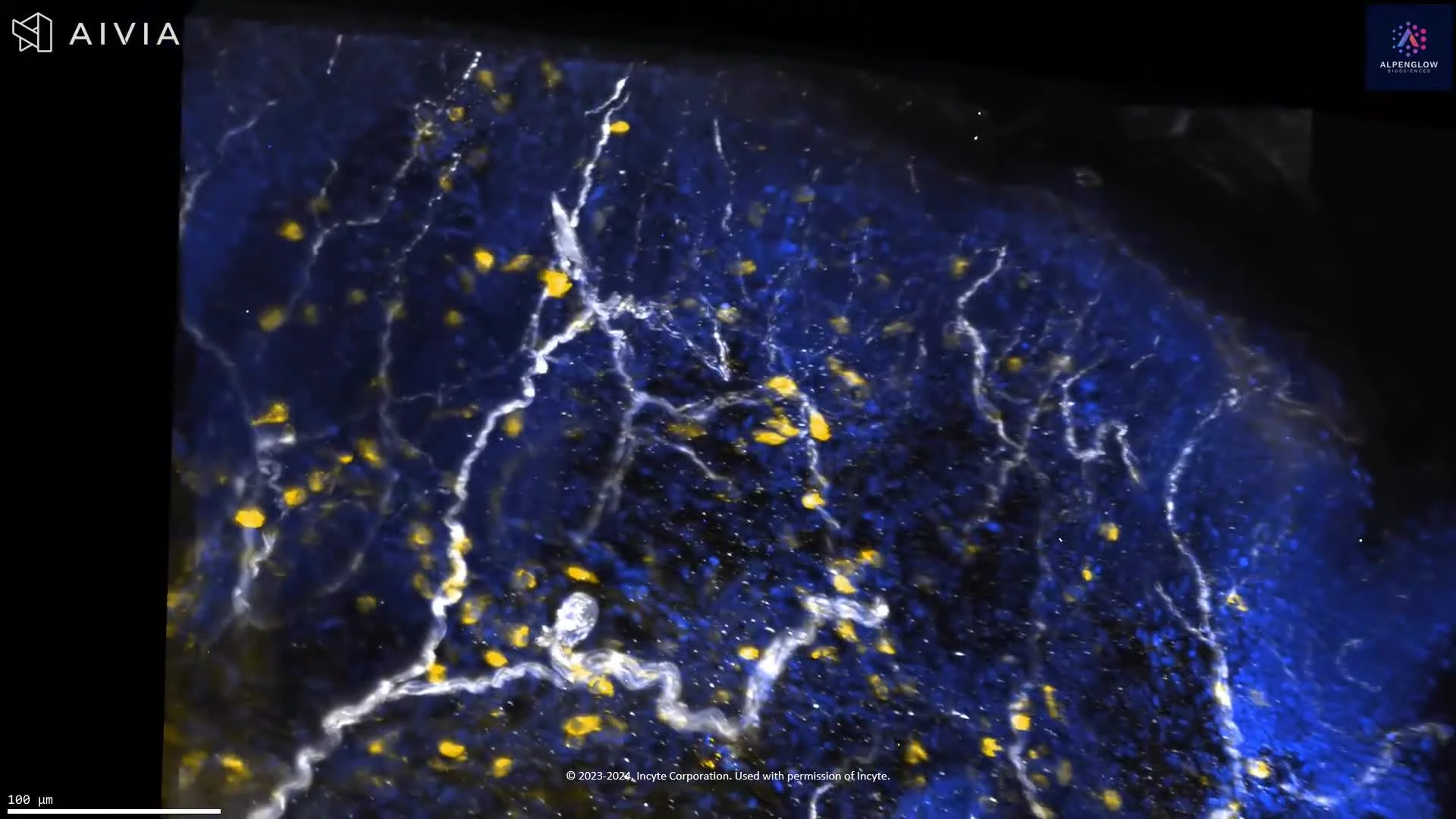
High-resolution 3D imaging of atopic dermatitis reveals precise innervation and immune cell distribution, surpassing conventional histology.
This high-resolution 3D visualization of an atopic dermatitis skin punch biopsy captures structural and cellular details that conventional tissue sections cannot achieve. Every nerve and immune cell is displayed in its true spatial context, offering clarity and depth into disease biology.
Stains used:
TO-PRO-3 (Blue): Highlights nuclear structures
PGP 9.5 (White): Maps precise innervation from larger nerves to finer branches
CD45 (Yellow): Clearly delineates immune cell distribution
Beyond visualization, the data is fully quantifiable through advanced data management workflows and AI-powered segmentation, enabling precise measurements of immune clustering around nerves.
This capability opens new avenues in dermatology research, driving insights into immune–neural interactions and inflammatory skin disease.
By combining whole-tissue 3D imaging with quantification, the dataset demonstrates the transformative potential of digital pathology and spatial biology.
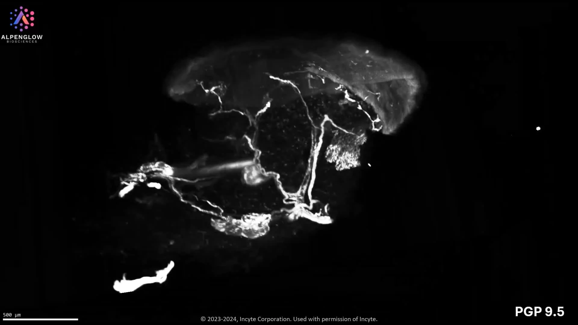
Journey through the delicate nerve structures of the epidermis and dermis at low resolution, and zoom in for an awe-inspiring close-up of lymphocyte distribution around these dynamic networks.
Immerse yourself in another breathtaking 3D skin punch biopsy, vividly stained to unveil:
PGP 9.5 (white) – tracing the intricate nerve networks
TO-PRO-3 (blue) – marking the nuclei
CD45 (yellow) – highlighting lymphocytes
Journey through the delicate nerve structures of the epidermis and dermis at low resolution, and zoom in for an awe-inspiring close-up of lymphocyte distribution around these dynamic networks. This visualization not only captures the stunning complexity of skin biology but also delivers actionable insights, propelling advancements in dermatology research.
See our AD use case.
40X image of a lesional Atopic Dermatitis samples stained with PGP9.5 (white) to highlight fine epidermal innervation, TO-PRO-3 (blue) for nuclear detail, and CD3 (green) to illuminate T-Cell infiltration of the Epidermis and Dermis.
High-resolution (40X) 3D imaging with Hybrid Open Top Light Sheet (HOTLS) microscopy of a Region of Interest in a lesional Atopic Dermatitis skin punch biopsy reveals intact tissue architecture and spatial immune organization.
The sample was stained with TO-PRO-3 for nuclei, PGP9.5 for nerve fibers, and CD3 for T cells, enabling detailed visualization of neuro-immune interactions and inflammatory pathways.
See our AD use case.
Image reproduced with permission from Incyte Corporation.
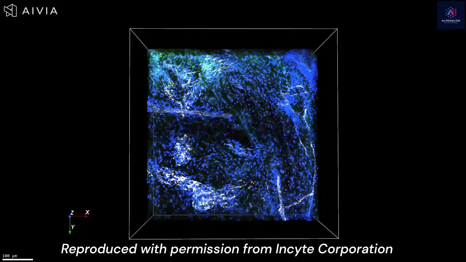
Low resolution image of a lesional Atopic Dermatitis sample stained with PGP9.5 (white) to highlight fine epidermal innervation, TO-PRO-3 (blue) for nuclear detail, and CD3 (green) to illuminate T-Cell infiltration of the Epidermis and Dermis.
Low-resolution 3D imaging with Hybrid Open Top Light Sheet (HOTLS) microscopy of an entire lesional Atopic Dermatitis skin punch biopsy highlights epidermal and dermal innervation alongside immune cell infiltration.
The tissue was stained with TO-PRO-3 (blue, nuclei), PGP9.5 (white, nerves), and CD3 (green, T cells), enabling comprehensive visualization of neuro-immune interactions and inflammatory changes across the full biopsy.
The tissue measured approximately 2.5mm X 2.5mm X 2.5mm.
See our AD use case.
Image reproduced with permission from Incyte Corporation.
Spatial dermatology readouts
Quantify skin structure, cells, and spatial relationships in 3D
Move beyond selected sections to examine how nerves, immune cells, vessels, follicles, and tissue compartments are organized across the full skin sample.
Skin architecture and remodeling
Examine epidermal and dermal organization, structural disruption, collagen-associated patterns, lesion boundaries, and localized tissue remodeling across the intact sample.
Peripheral innervation and neuroimmune biology
Trace nerve fibers, branching patterns, epidermal innervation, and the spatial proximity of nerves to immune cells across the tissue volume.
Immune infiltration and cellular organization
Quantify immune density, distribution, clustering, and localization across skin layers, lesions, follicles, vessels, and other tissue compartments.
Follicles, vasculature, and treatment response
Assess hair follicles, pilosebaceous structures, microvascular organization, and treatment-associated changes across experimental groups or disease stages.
Next step
Bring your skin biology question into 3D
Share your tissue type, disease model, marker strategy, and intended spatial readouts. Alpenglow can help define an intact-tissue imaging and quantitative analysis approach for your dermatology study.
Explore how 3D Derm Score™ supports quantitative assessment of nerves, immune cells, follicles, vessels, and tissue architecture.
Discuss your dermatology study
Tell the Alpenglow team which skin structures, cellular populations, and spatial relationships you need to visualize or quantify.

3D fluorescence imaging of mouse skin reveals the continuity and branching of sensory nerve networks across tissue depth. PGP9.5 highlights broader nerve architecture, while the Nav1.8-associated signal identifies a subset of sensory neurons.
This dataset presents mouse skin tissue imaged in 3D using fluorescence labeling. PGP9.5 highlights the broader nerve network, YO-PRO-1 labels nuclei, and Nav1.8-associated signal identifies a subset of sensory neurons.
The volumetric view preserves the continuity of nerve fibers across the imaged tissue depth, revealing large nerve bundles, finer branches, crossings, and terminal structures within dermal and epidermal regions.
With appropriate segmentation, the dataset can support measurement of nerve density, fiber length, branching, orientation, and spatial relationships with surrounding cells and tissue structures. These features are relevant to research on cutaneous innervation, sensory biology, pain, itch, inflammation, and treatment-associated changes in nerve architecture.
The tissue was imaged using the Aurora 3D™ Spatial Biology Solution, including the 3Di™ Hybrid Open-Top Light-Sheet microscope.