3D imaging of nerve innervation at the dermal–epidermal junction
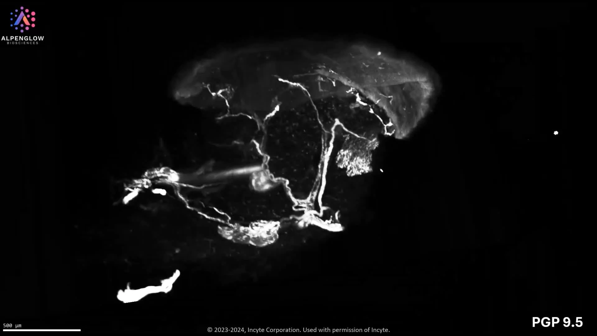
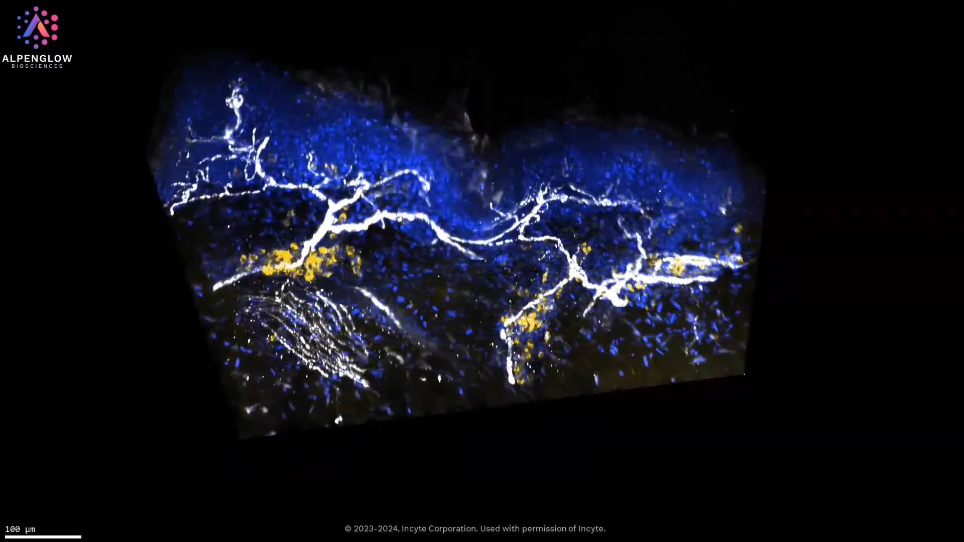
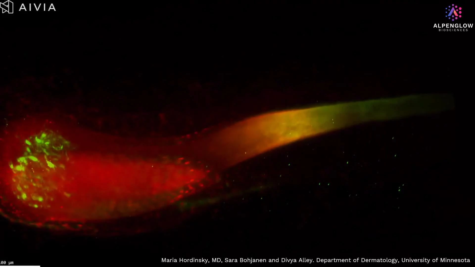
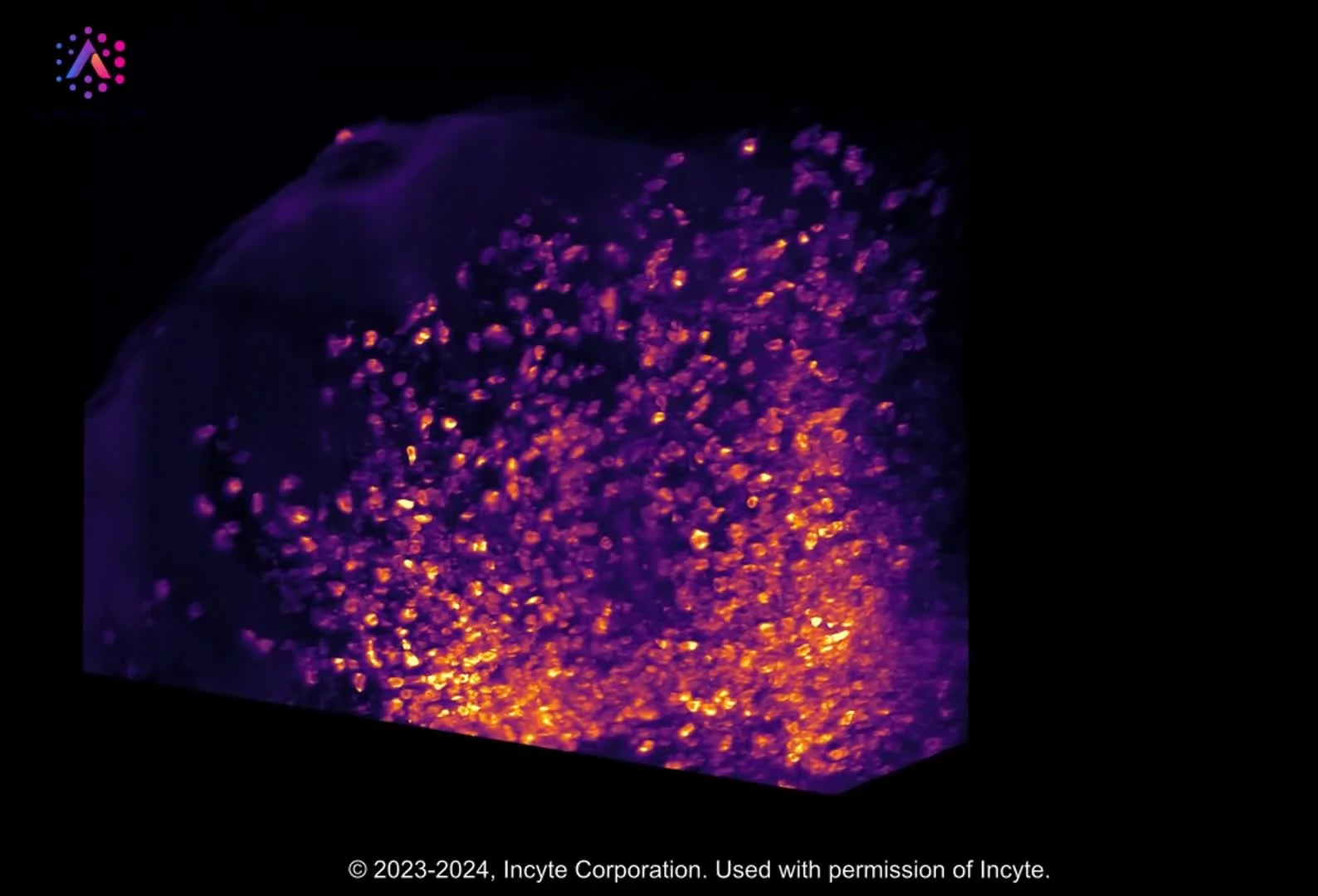
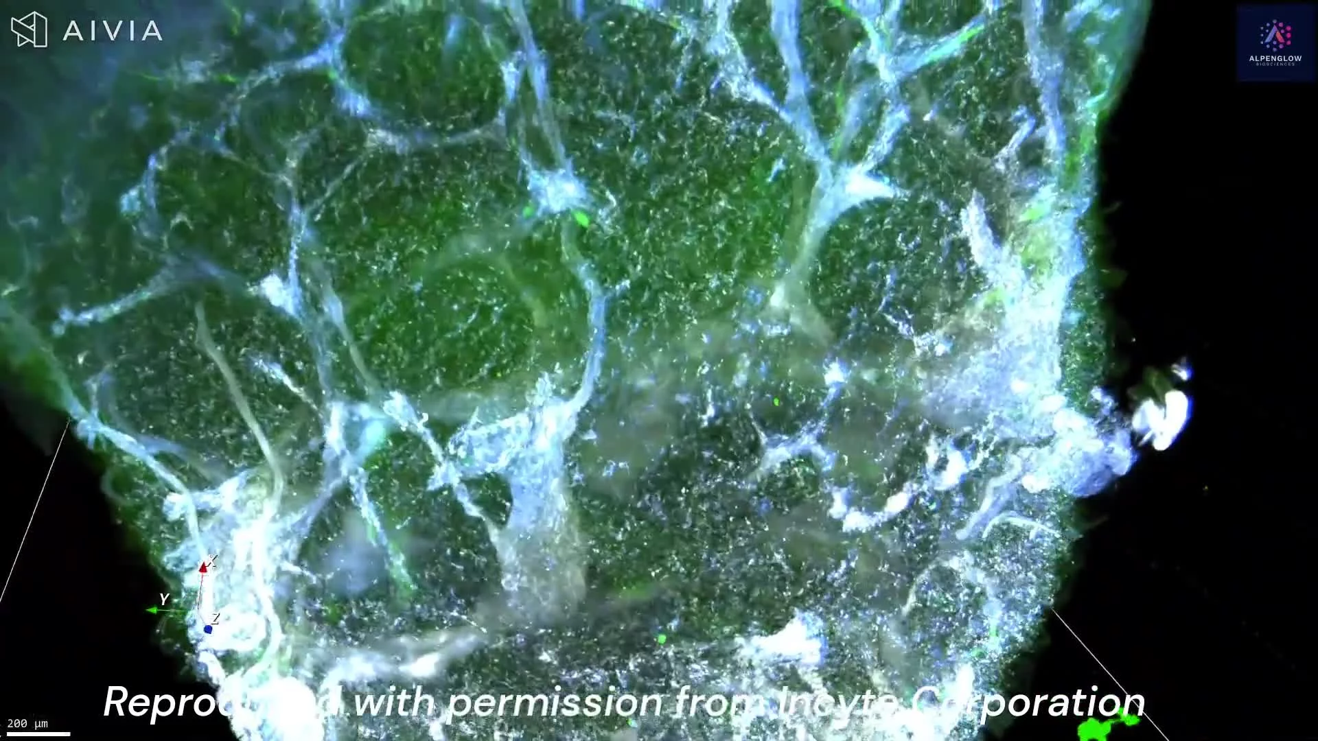
This dataset presents high-resolution 3D tissue imaging of rat toe pad skin, focusing on nerve innervation at the dermal–epidermal junction. Using Hybrid Open-Top Light-Sheet microscopy, intact tissue is imaged volumetrically to preserve full structural continuity.
Nerves are labeled with PGP9.5 (white) and nuclei with YoPro-1 (blue), revealing dense innervation patterns, complex branching, and crossings extending across tissue depth. From large nerve bundles to fine terminal fibers, the architecture of the peripheral nervous system is captured without disruption.
This approach enables quantitative tissue analysis and spatial profiling of nerve networks in their native context, supporting studies of skin biology, neuro-immune interactions, and disease mechanisms. By moving beyond thin sections, 3D imaging provides a more complete understanding of tissue organization and cellular relationships.