
Pathologist
3D digital pathology for intact tissue
Extend pathology beyond the selected section
Preserve whole-tissue architecture and examine margins, glands, immune structures, vessels, nerves, and rare features across the full sample volume. Alpenglow combines non-destructive 3D tissue imaging with AI-powered analysis to generate volumetric evidence for research pathology, spatial biomarker studies, and translational workflows.
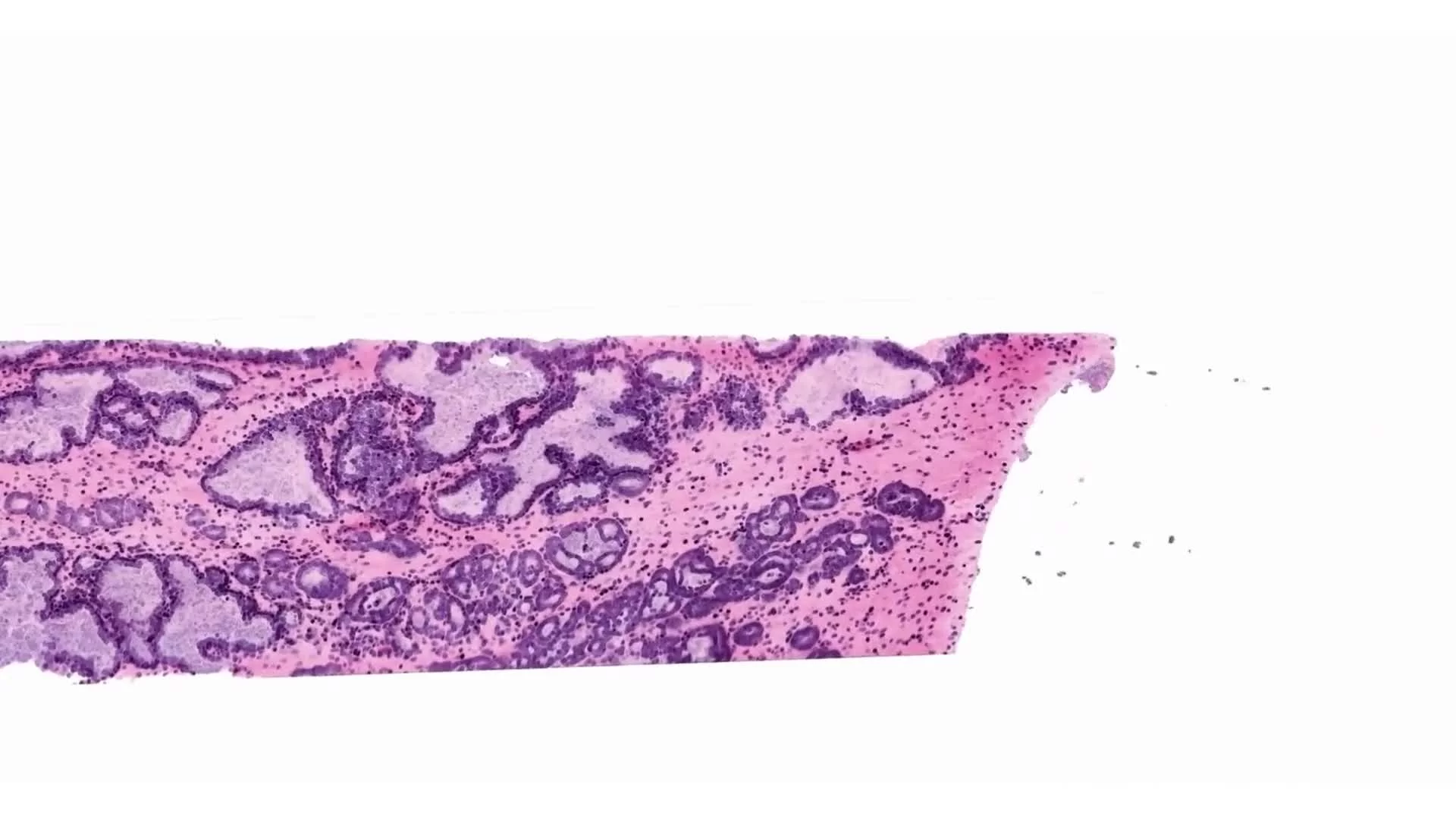
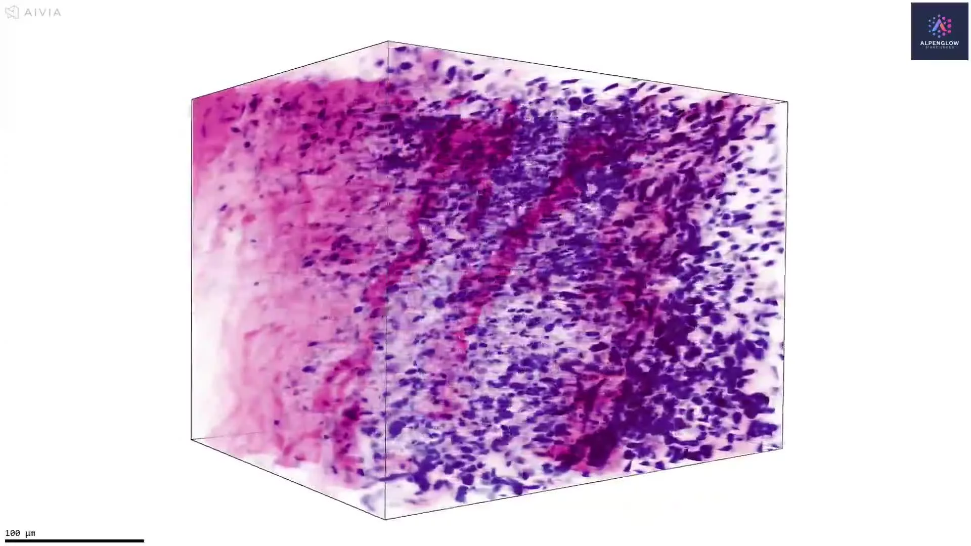
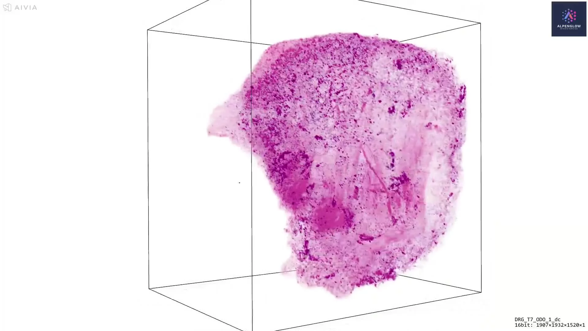
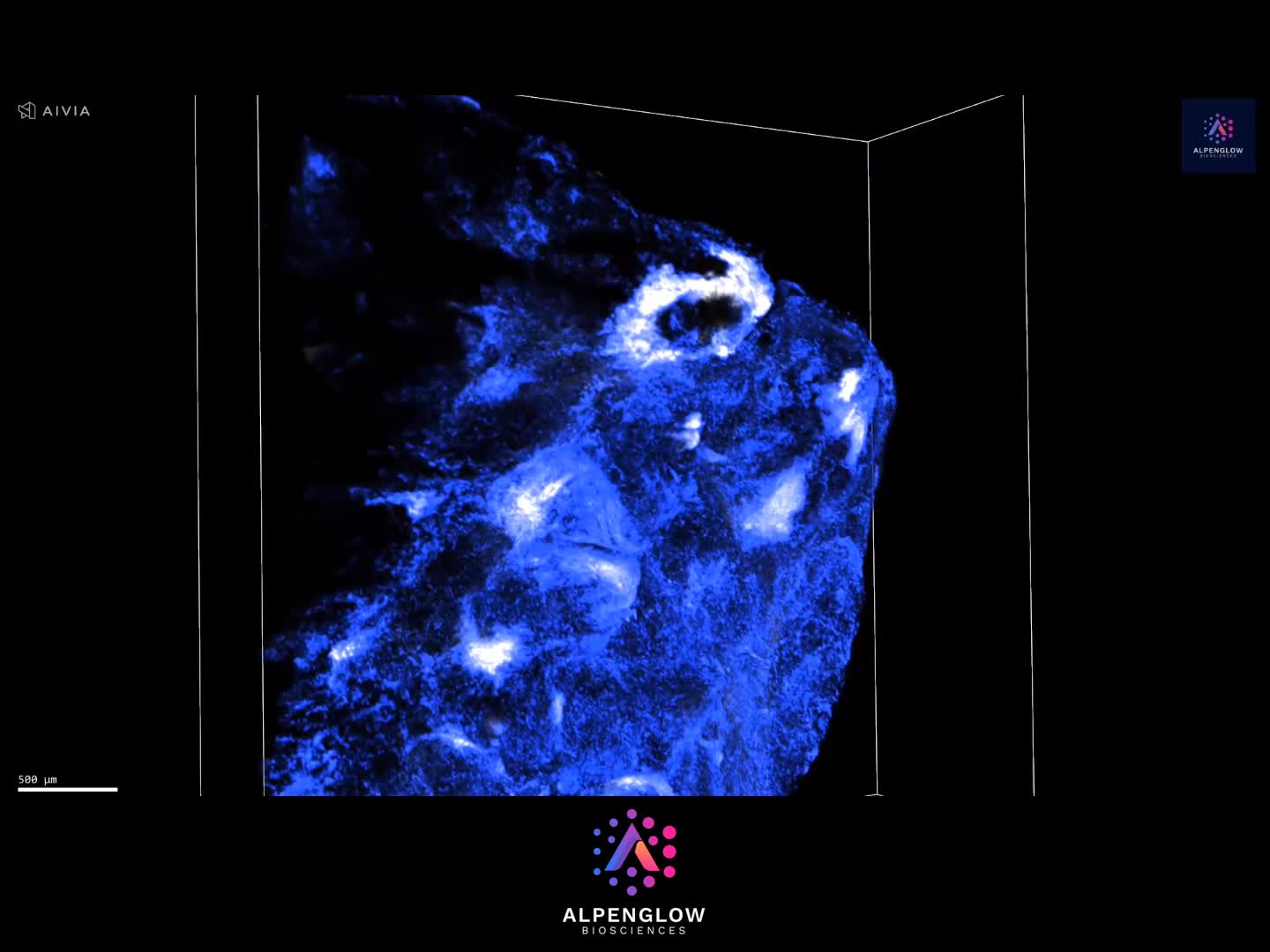

Volumetric pathology readouts
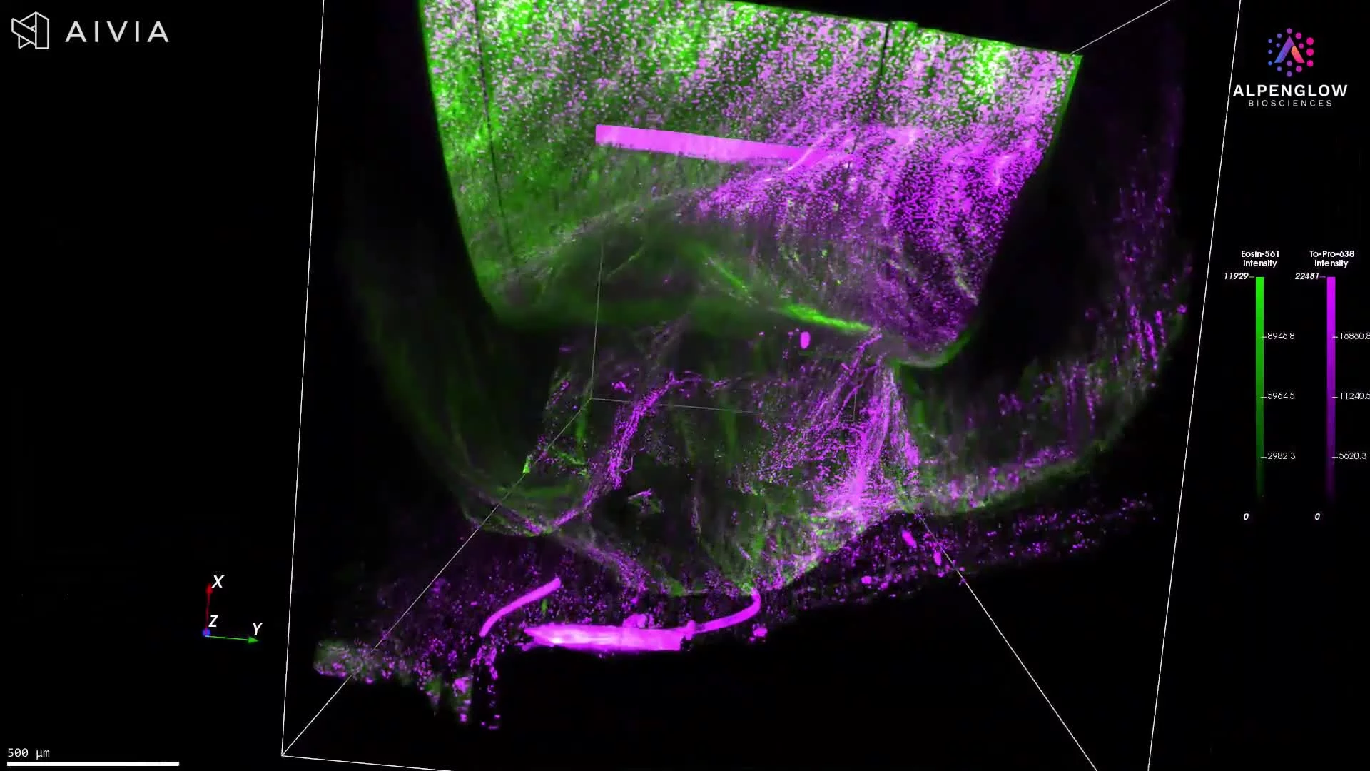
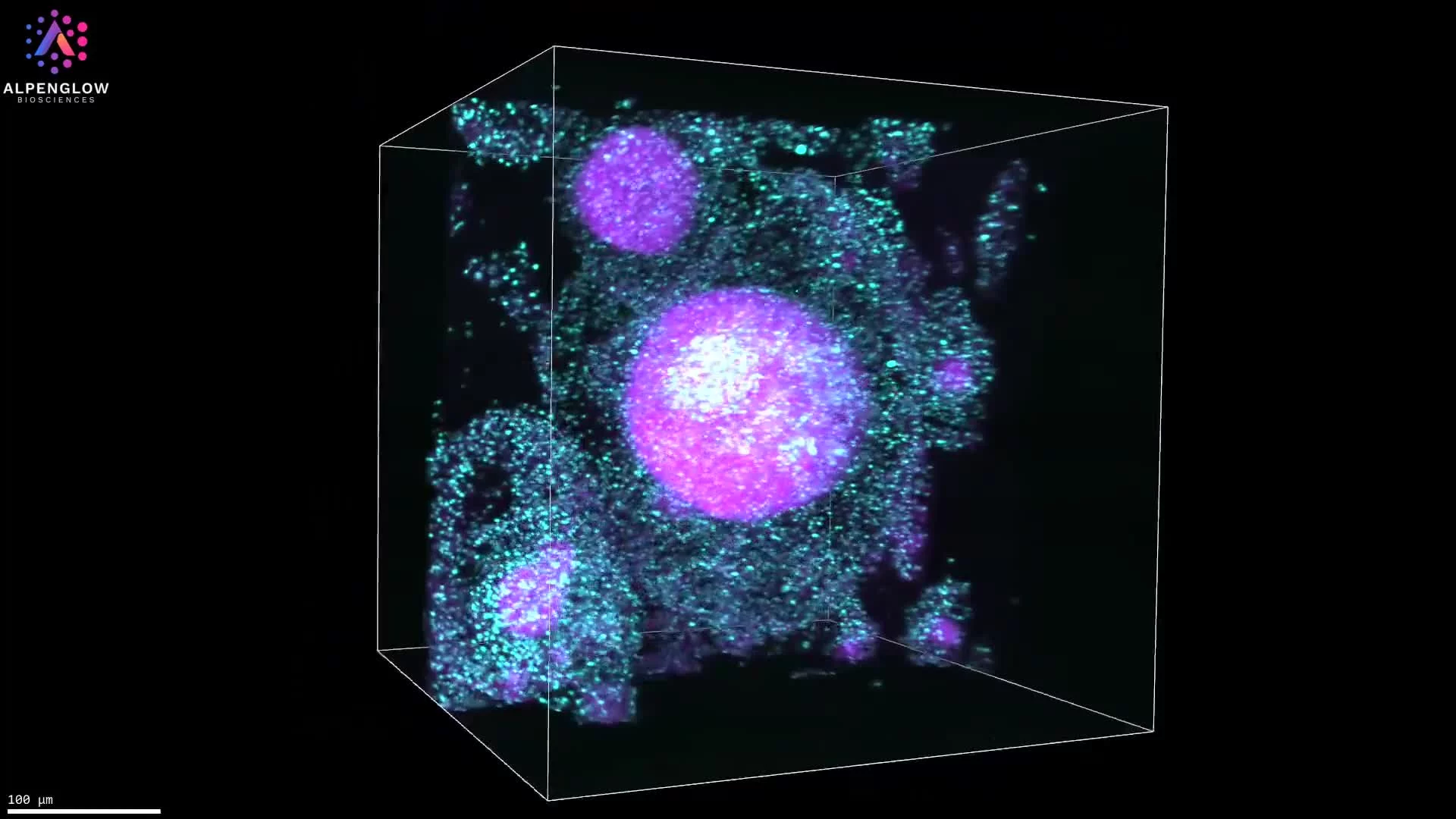
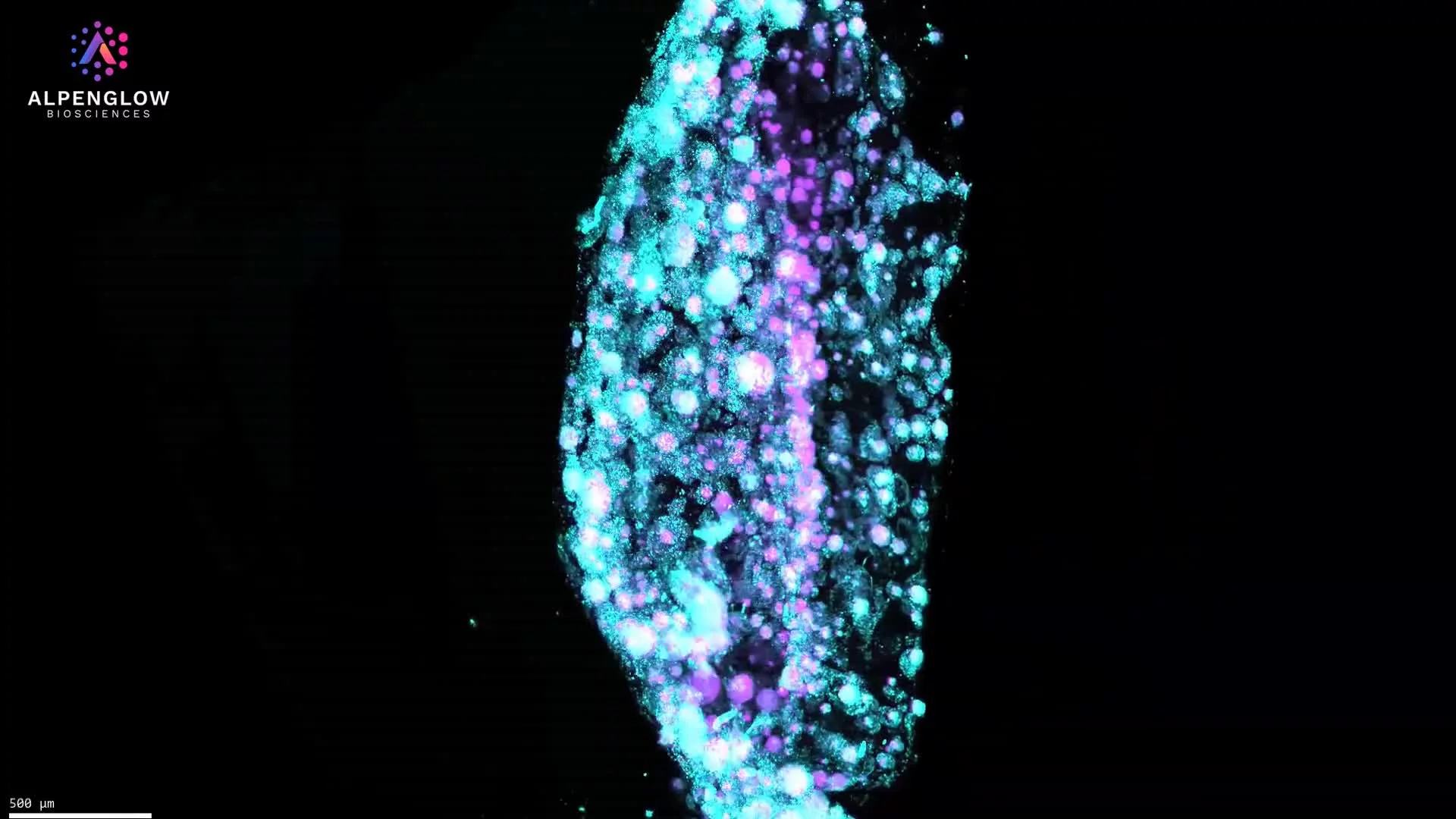
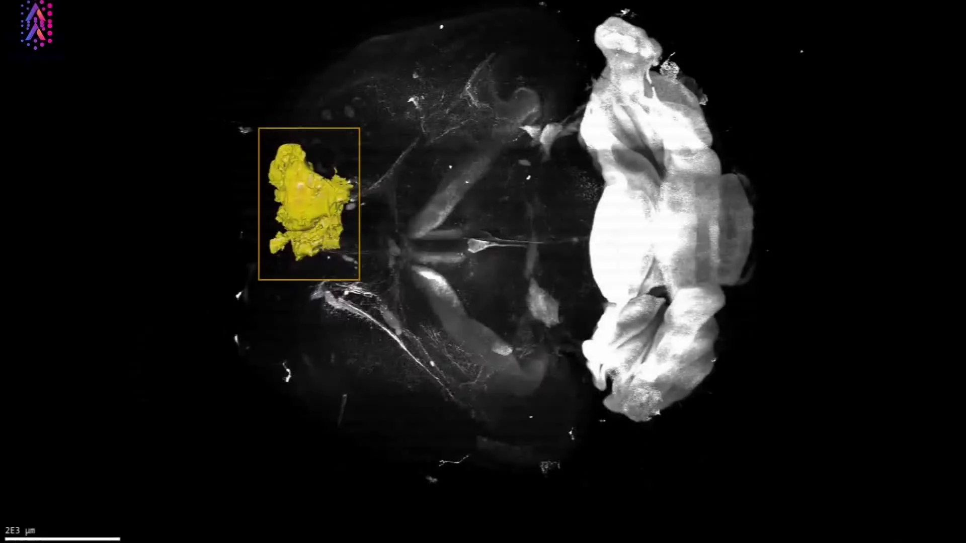
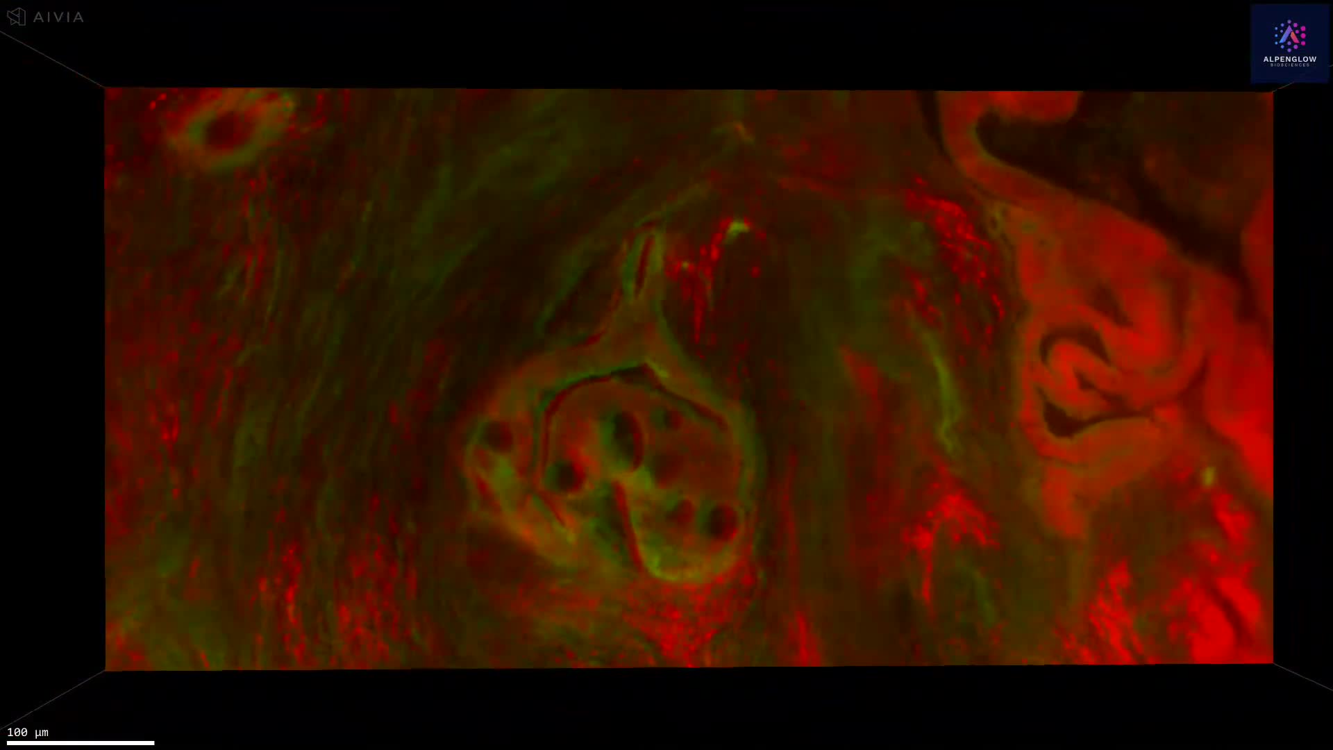
Examine pathology features across depth, not one plane
Extend tissue review beyond selected sections to evaluate biological features continuously across the sample. Intact 3D imaging preserves morphology, spatial relationships, and tissue-scale organization for research pathology and digital pathology programs.
Broader tissue sampling
Examine more tissue volume across regions and depths, reducing reliance on a small number of selected planes when studying heterogeneous or unevenly distributed biology.
Published example
Whole-lymph-node OTLS enabled volumetric assessment of breast cancer metastases, revealing metastatic extent that could be underestimated from selected 2D sections.
Read the Journal of Biomedical Optics studyBoundaries and structural continuity
Follow tissue regions, lesion boundaries, glands, vessels, nerves, and branching structures continuously through the volume rather than reconstructing them from disconnected fragments.
Published example
GlaSkeN used OTLS to preserve complete prostate gland networks and quantify branching, curvature, torsion, and connectivity. 3D architectural features were associated with biochemical recurrence across two cohorts.
Read the Modern Pathology studyRare and spatially restricted features
Locate focal lesions, sparse cells, localized immune structures, and other features that may occur outside a selected 2D section.
Published example
TRICARE analyzed complete 3D pathology datasets to identify high-risk tissue levels for pathologist review in prostate biopsies and Barrett's esophagus specimens.
Read the Nature Biomedical Engineering studyQuantitative marker and spatial review
Measure cells, structures, regions, marker distributions, and spatial relationships across intact tissue to support research, biomarker development, reference datasets, and computational pathology workflows.
Published example
SIGHT used generative immunolabeling to automate delineation of benign and prostate-cancer-enriched regions within 3D pathology datasets.
Read the Modern Pathology studyNext step
Bring your pathology question into 3D
Share your tissue type, structures of interest, marker strategy, and intended quantitative readouts. Alpenglow can help define a non-destructive 3D histology workflow for research pathology, biomarker studies, and computational pathology programs.
Building AI or diagnostic reference datasets? Explore ground-truth 3D tissue data for volumetric imaging, expert annotation, and quantitative tissue measurements.
Discuss your pathology study
Tell the Alpenglow team which tissue features, spatial relationships, and measurements you need to review across the intact sample.

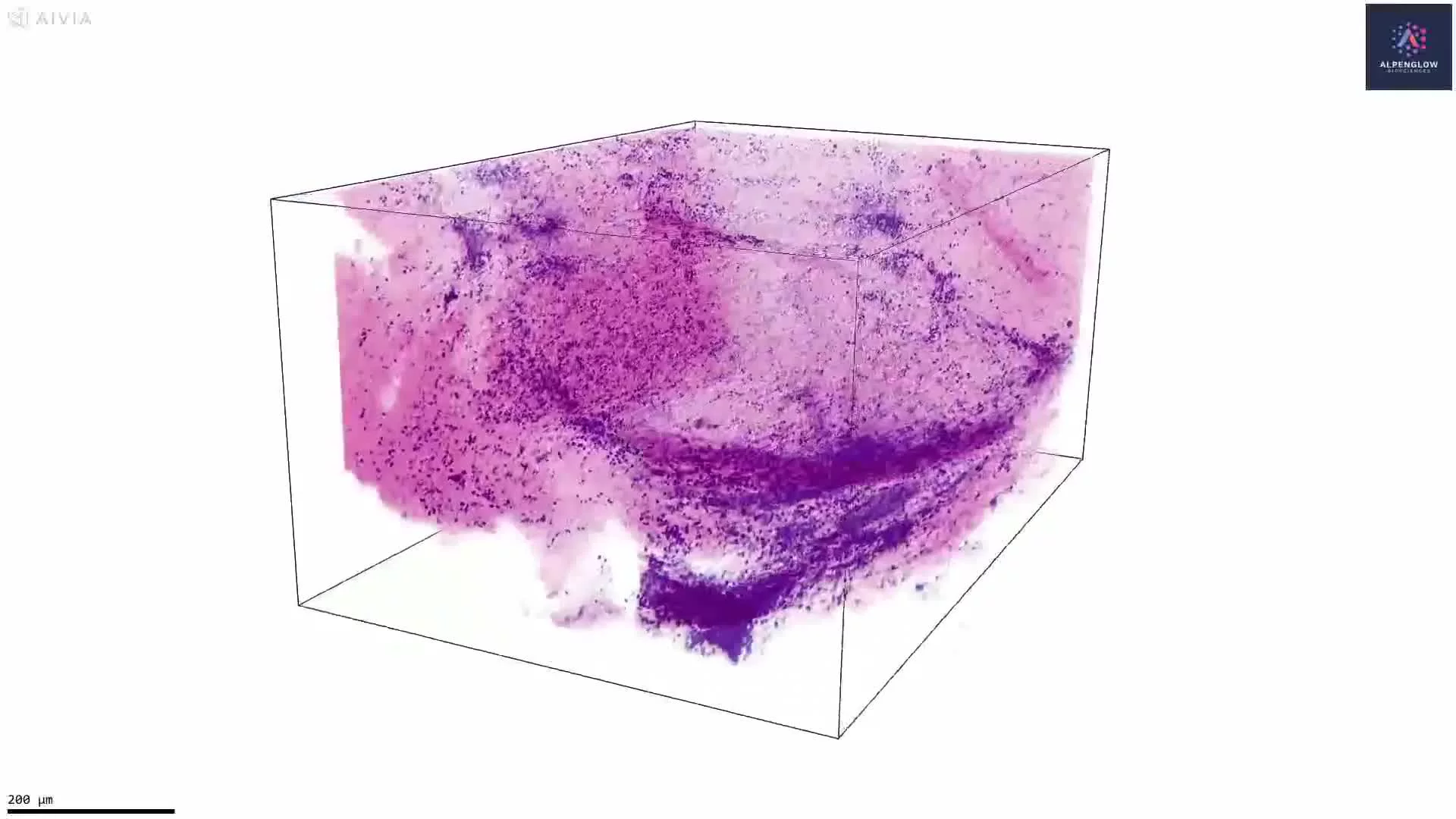
Explore human duodenum tissue in 3D using eosin and TO-PRO-3 in an H&E-like visualization. Volumetric imaging preserves villous morphology and spatial organization across depth and can support quantitative analysis of villus dimensions, density, spacing, and regional variation.
This video presents a volumetric visualization of human duodenum tissue stained with eosin and the nuclear marker TO-PRO-3, then pseudocolored to create an H&E-like appearance.
3D tissue imaging reveals intestinal villi as continuous structures across the imaged volume, preserving information about their morphology, orientation, spacing, density, and regional organization. Viewing villi across depth also reduces the influence of sectioning angle and sampling location that can affect measurements from individual 2D sections.
With appropriate segmentation, the dataset can support quantitative analysis of villus number, height, width, volume, elongation, spacing, density, and variation across tissue regions.
These measurements are relevant to gastrointestinal research, including studies of celiac disease, where villous architecture may become shortened, flattened, fused, or otherwise disrupted. Quantifying these changes across a tissue volume could help characterize the distribution and heterogeneity of structural alterations.
The tissue was imaged on the Aurora 3D™ platform using the 3Di™ Hybrid Open-Top Light-Sheet (HOTLS) microscope.