
Research Group Leader
3D spatial biology for research leadership
Build a 3D imaging strategy your research group can scale
Equip your team with a connected workflow for intact tissue imaging, volumetric data management, and AI-powered analysis. Alpenglow helps research groups standardize complex 3D studies, support diverse tissue models, and generate quantitative spatial readouts across exploratory, translational, and collaborative programs.
A shared research capability
Turn 3D imaging into a strategy for the whole group
Create a consistent approach to intact tissue imaging, spatial profiling, and quantitative analysis across projects. A connected workflow helps teams compare studies, manage complex volumetric data, and build reusable imaging and analysis methods.
Standardize methods across projects
Establish repeatable imaging, data processing, and quantitative analysis workflows that can be applied across researchers, experimental conditions, and longitudinal studies.
Support diverse biological models
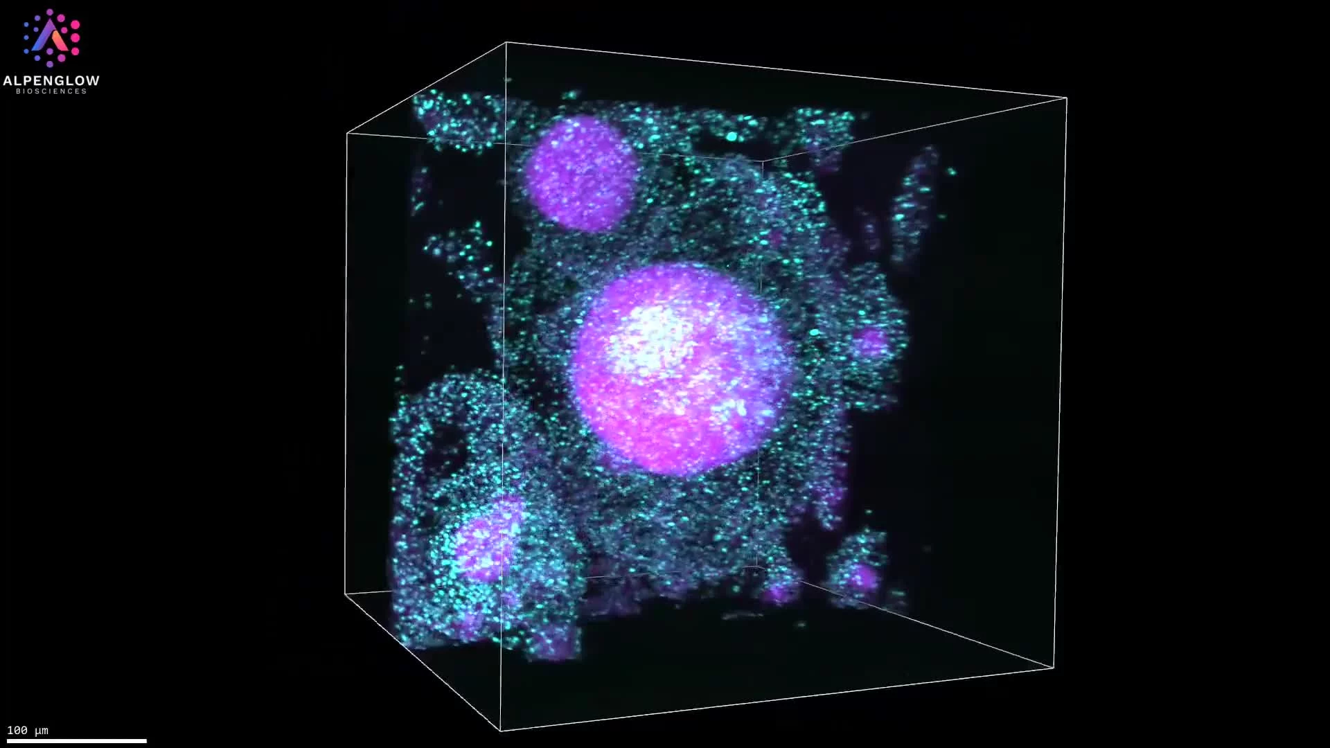
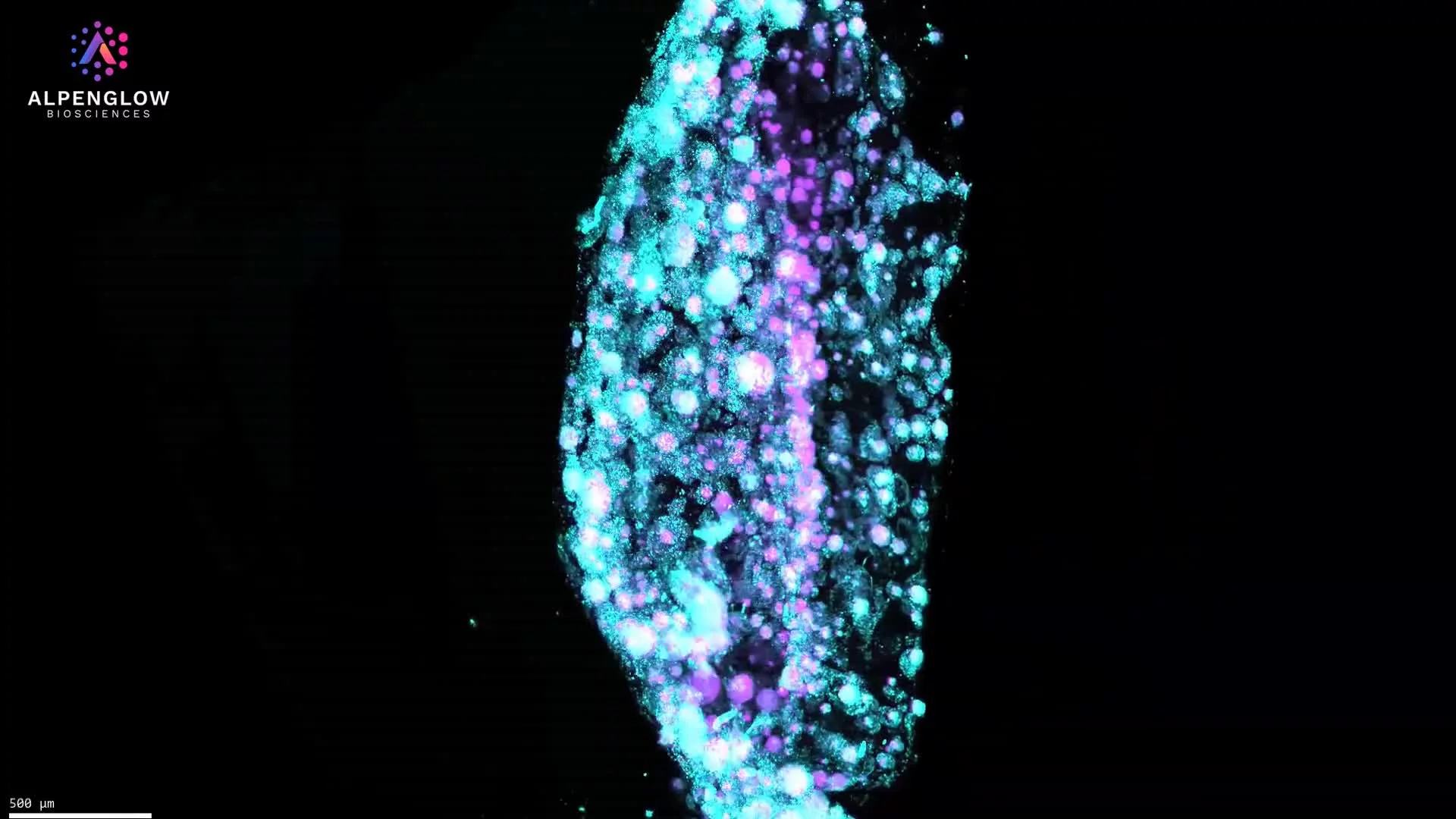
Apply intact 3D tissue imaging across organoids, tissue blocks, preclinical models, whole organs, and other complex specimens without limiting the group to one sample format or application.
Scale from pilots to larger studies
Begin with focused feasibility studies, then extend validated staining, imaging, and analysis methods across larger sample sets and collaborative research programs.
Strengthen collaborative research
Generate quantitative volumetric datasets, interactive visualizations, and defined spatial readouts that can support collaboration, publication, grant development, and shared interpretation across disciplines.
Build the strategy
Define the right 3D imaging approach for your research program
Share your scientific priorities, tissue models, team structure, and intended quantitative readouts. Alpenglow can help assess how intact 3D tissue imaging, spatial profiling, and AI-powered analysis could support current studies and future program growth.
Explore the connected Aurora 3D™ workflow for imaging, volumetric data management, and quantitative analysis.
Discuss your research program
Review your sample types, scientific questions, workflow requirements, and plans for scaling 3D imaging across the group.

From vasculature to single cells, 3D light-sheet imaging reveals the intricate architecture of a whole mouse brain in unparalleled detail.
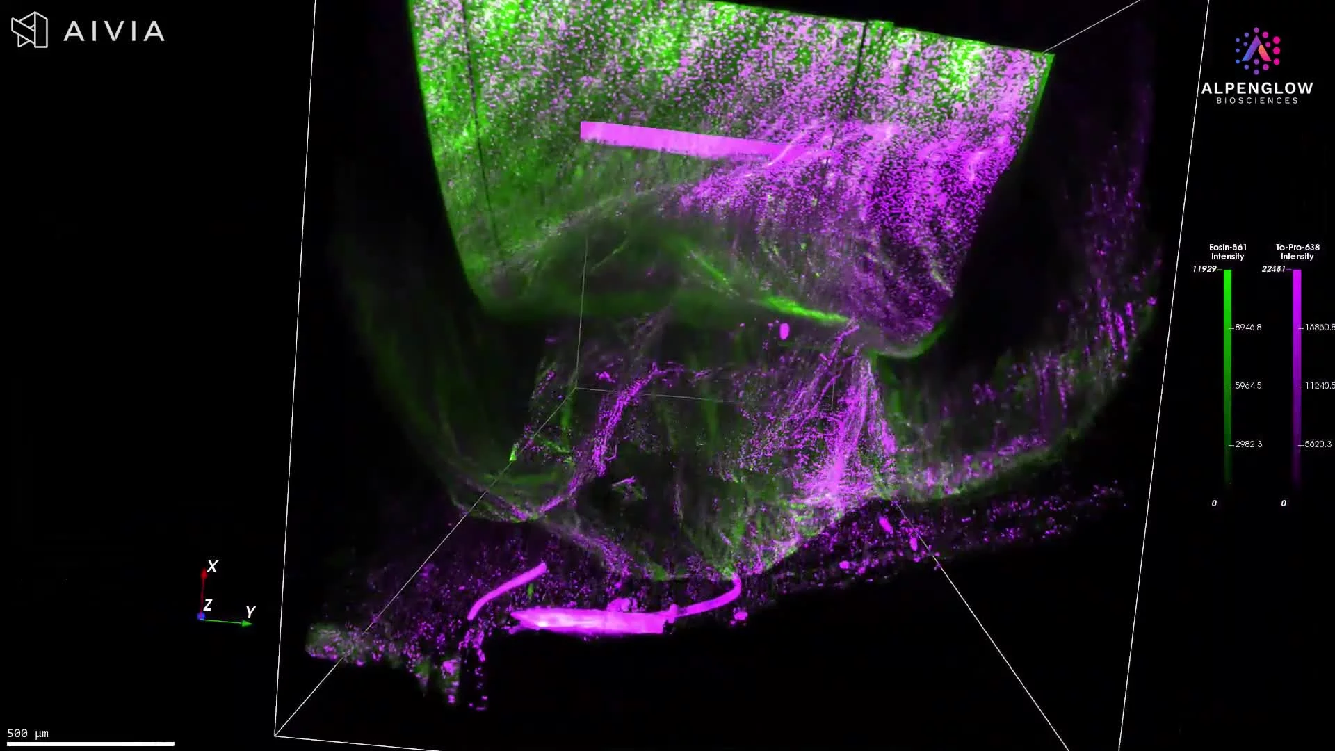
This dataset showcases 3D light-sheet imaging of a whole mouse brain, captured with the Aurora™ 3Di Hybrid Open Top Light Sheet (HOTLS) microscope. From organ-level vasculature down to single-cell resolution, the imaging reveals the full intricacy of brain architecture across every layer.
The dataset highlights how volumetric imaging preserves intact morphology, making it possible to study neuronal networks, vascular pathways, and cellular organization without the distortions of traditional 2D histology.
Why 3D over 2D?
Complete visualization of neuronal and vascular networks across the brain
Cellular context preserved in full volumetric detail
Accurate mapping of tissue complexity that 2D slices cannot replicate
This dataset connects directly to the Nature Methods paper presenting our light-sheet microscope: Read the paper.
By combining 3Dm data management with 3Dai AI-powered segmentation, researchers can extend these visuals into actionable analyses, from neuronal density measurements to vascular branching and spatial profiling.
Applications include neuroscience research, brain mapping, and studies of neurodegenerative diseases, where understanding tissue complexity in situ is critical.