3D Tissue Imaging of Mouse Muscle Innervation
This dataset showcases high-resolution 3D tissue imaging of mouse muscle, designed for quantitative spatial biology and digital pathology applications.
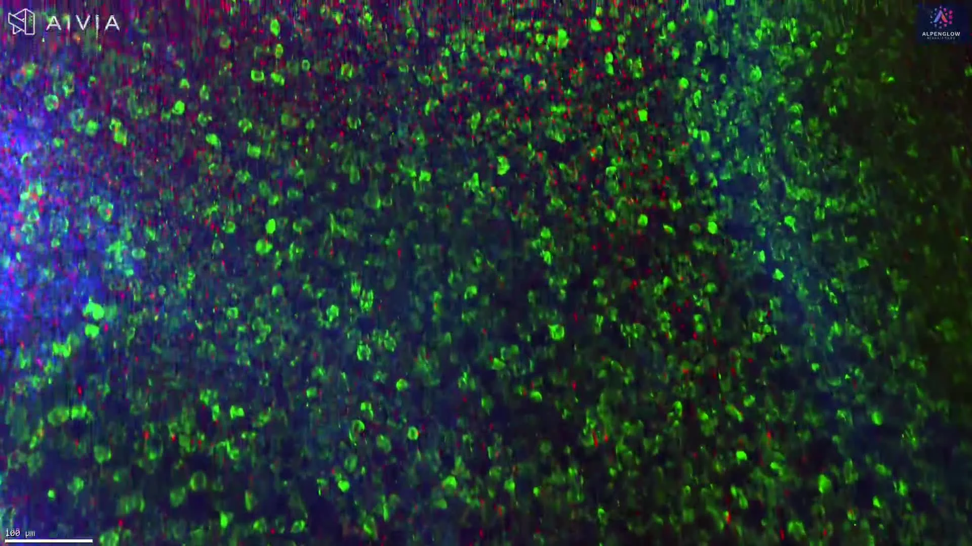
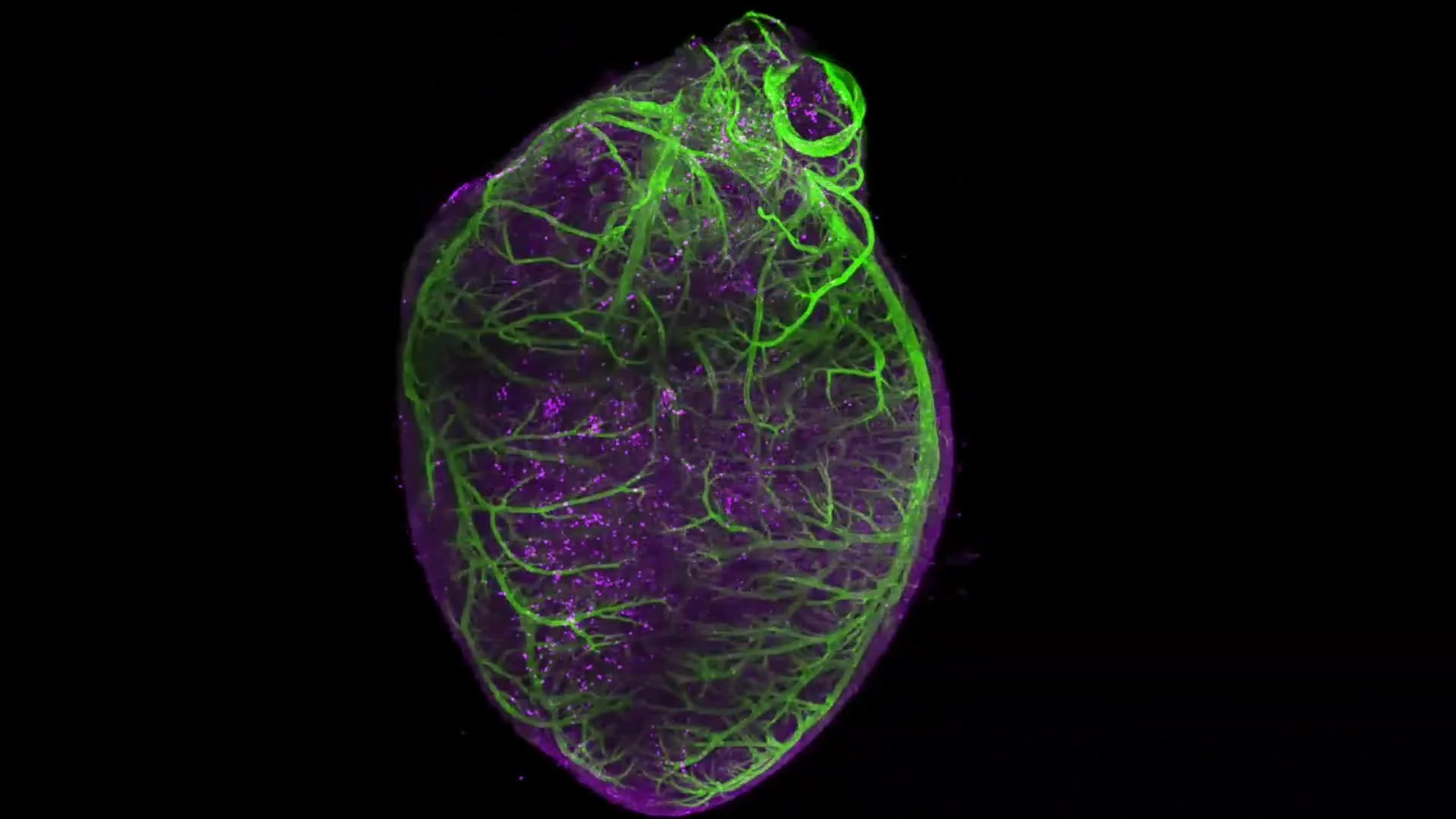
NF200 (green) labels neurofilament heavy chain in large-caliber axons, frequently associated with myelinated fibers, enabling visualization of major neural trajectories.
PGP9.5 (magenta), a widely used neuronal marker, maps the broader axonal network interlacing the muscle.
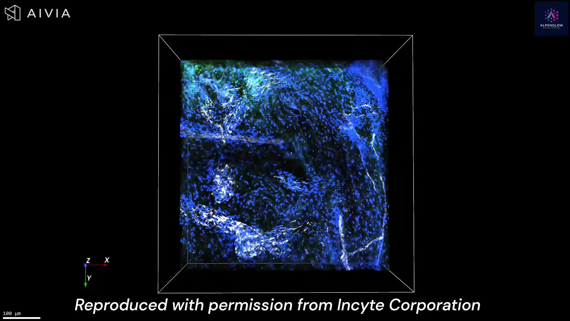
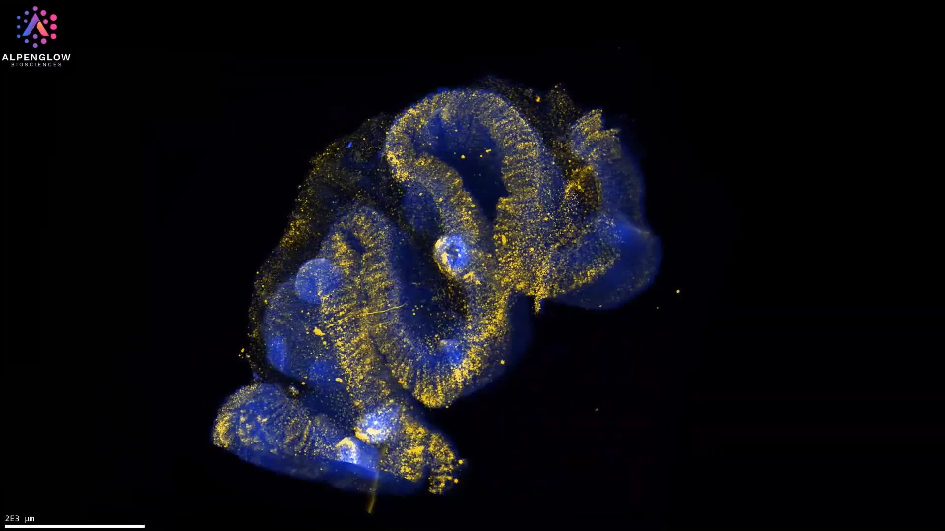
YO-PRO-1 (blue) stains nuclei, providing a consistent cellular reference throughout the volume.
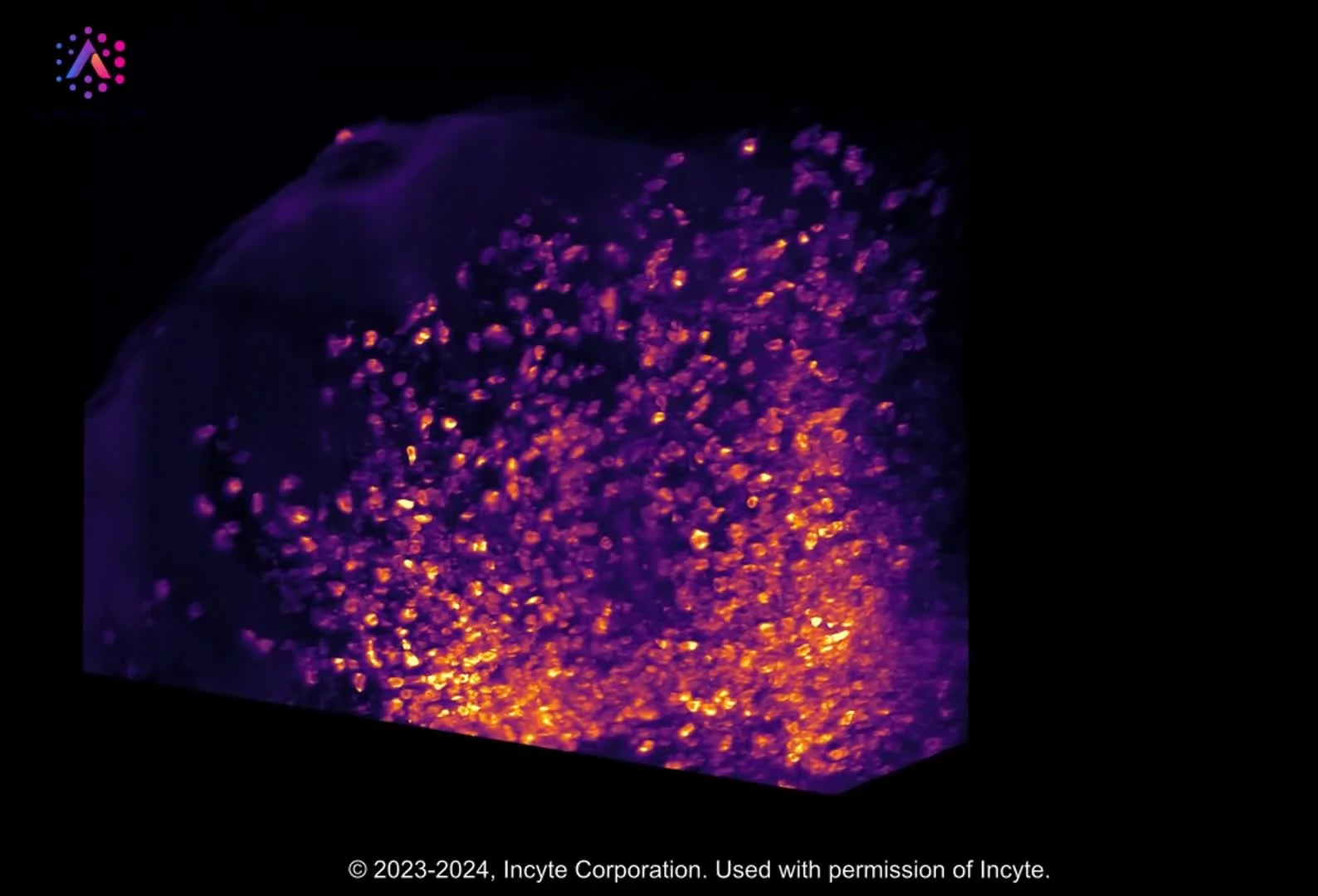
Volumetric imaging preserves branching hierarchies, axonal trajectories, and regional innervation patterns that are fragmented in thin sections. Entire arborization structures can be followed through depth, allowing measurement of axon length, branching complexity, regional density, and spatial distribution.
When combined with AI-powered analysis, this approach supports quantitative tissue analysis, tissue microenvironment visualization, and spatial profiling at scale. The result is high-resolution 3D imaging that converts structural complexity into measurable biological insight.