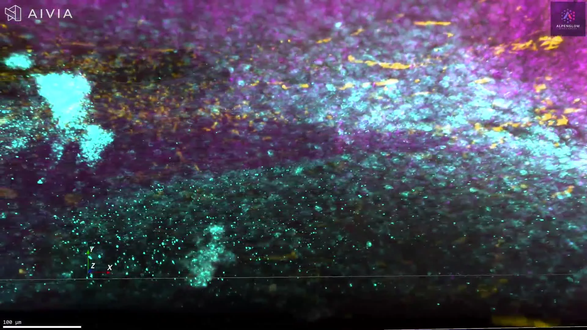
Expanded 3D imaging of mouse kidney. Glomerular and extracellular matrix architecture at subcellular scale
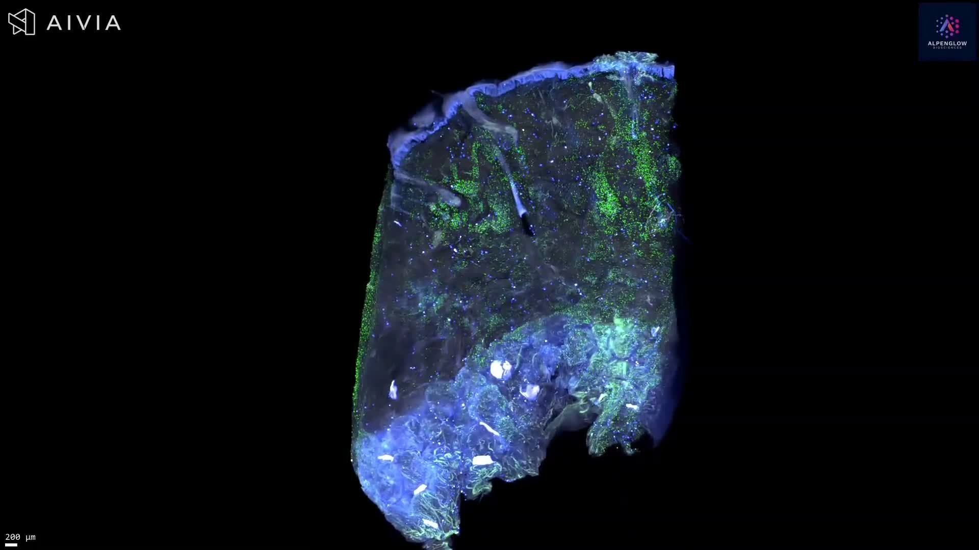
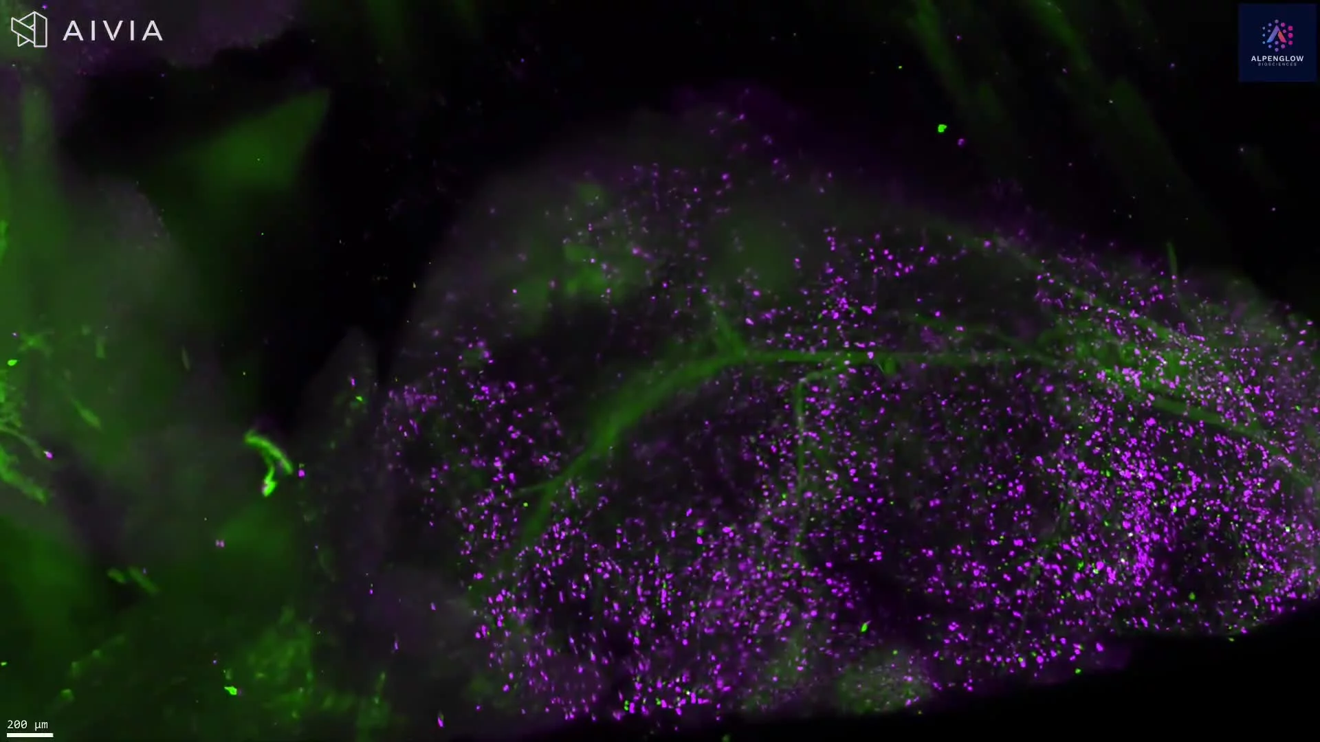
This video presents expanded 3D imaging of mouse kidney tissue, revealing glomerular, basement membrane, and extracellular matrix architecture across an intact tissue volume.
Nuclei are shown in white. Collagen IV, shown in blue, highlights basement membrane structures. WGA lectin, shown in magenta, labels glycoprotein-rich tissue features, while podocalyxin, shown in green, highlights podocyte-associated structures within the glomeruli.
Expansion microscopy physically enlarges the specimen, increasing the separation between fluorescently labeled structures while preserving their relative spatial organization. Traversing the expanded volume reveals the three-dimensional relationships among glomeruli, basement membranes, and surrounding renal tissue that can appear fragmented in individual 2D sections.
The dataset can support analysis of glomerular morphology, basement membrane organization, spatial distribution, and structural variation across the imaged kidney volume.
This work was performed in collaboration with Joshua Vaughan and the University of Washington Department of Chemistry.
The tissue was imaged using the Aurora 3D™ Spatial Biology Solution, including the 3Di™ Hybrid Open-Top Light-Sheet microscope.