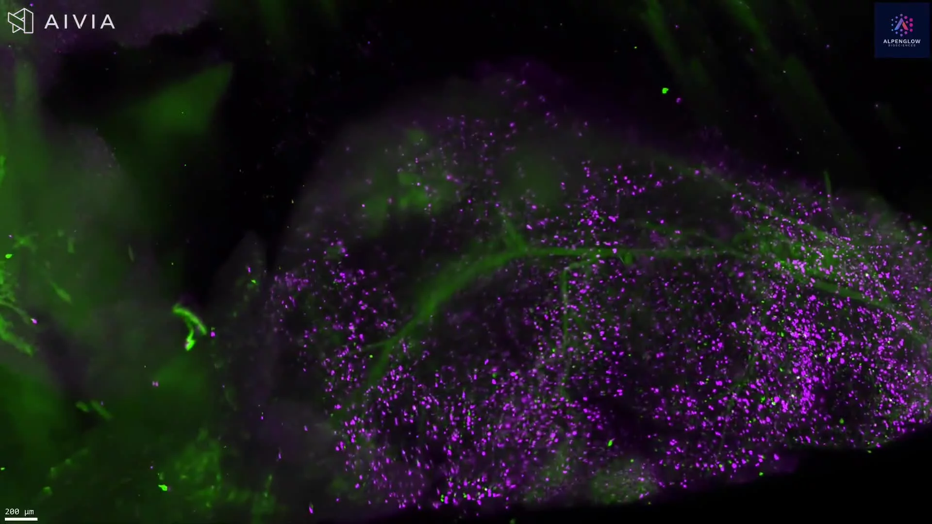
3D Imaging of Vascular, Macrophage, and Nerve Architecture in Cleared Mouse Fat Pad
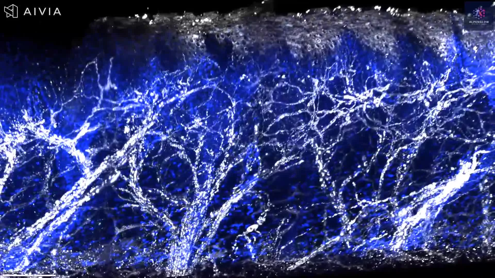
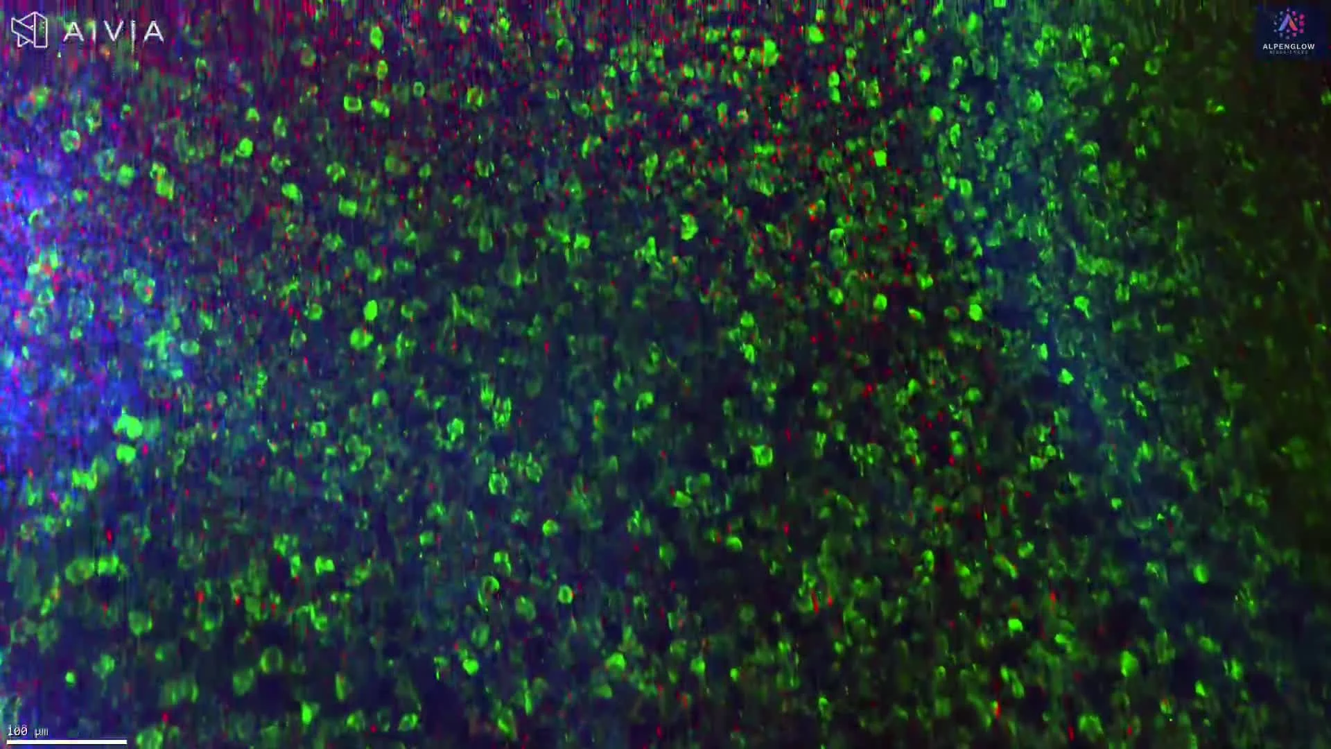
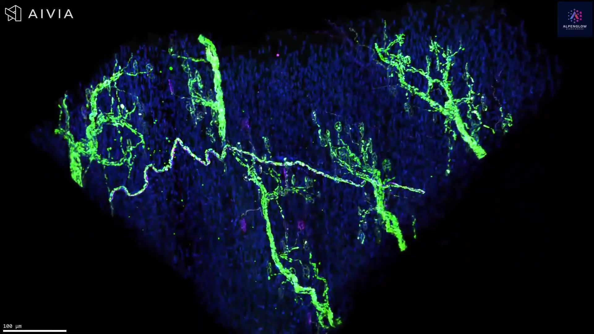
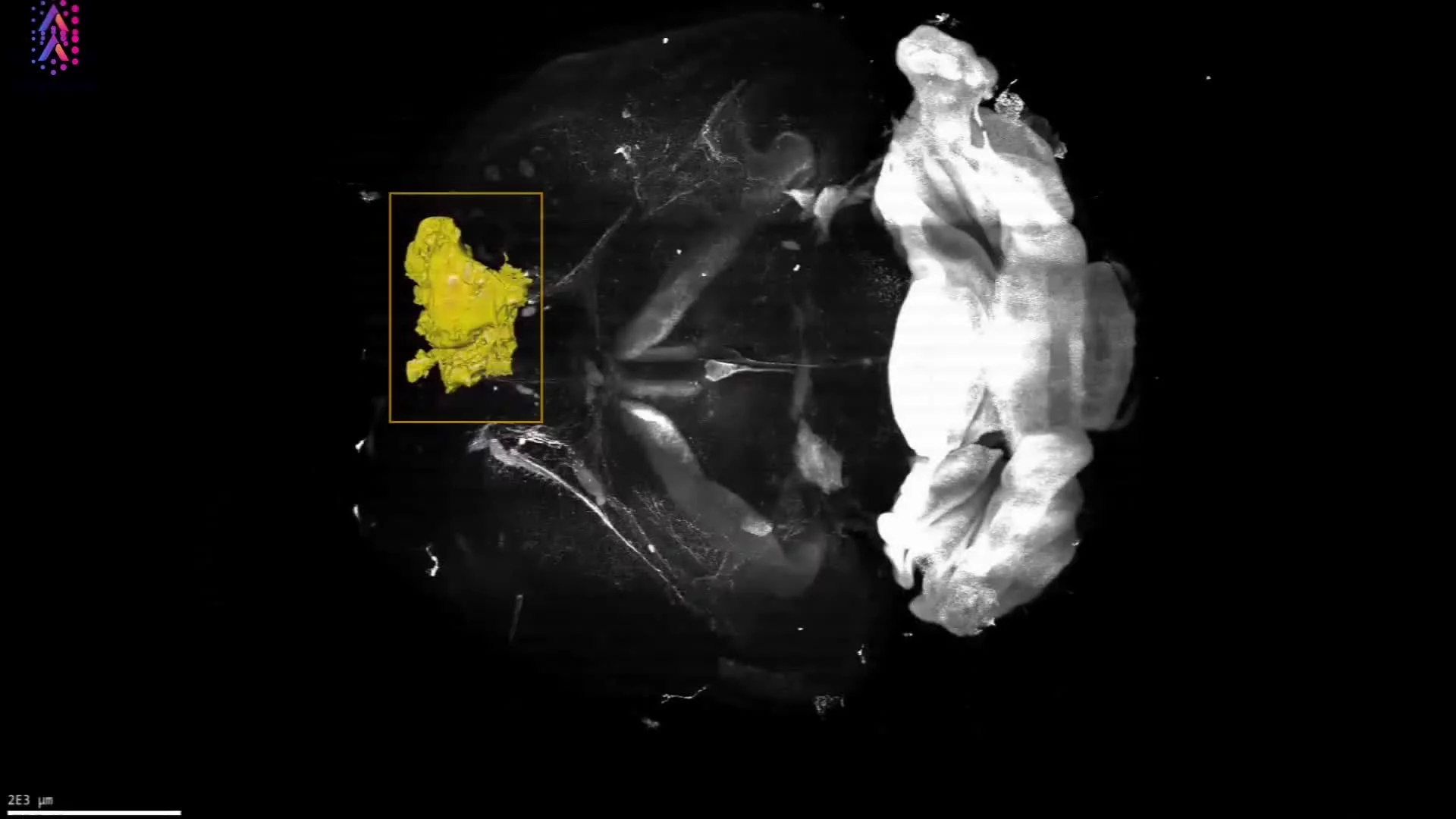
This dataset presents a cleared, triple-labeled mouse fat pad imaged in 3D, preserving vascular, immune, and neural structures across the intact tissue volume.
Lectin, shown in red, highlights the vascular network. CD68, shown in turquoise, identifies macrophage-associated cells, while PGP9.5, shown in green, traces nerve fibers throughout the adipose tissue.
The volumetric view reveals the distribution and spatial relationships of blood vessels, CD68-positive cells, and nerves within the surrounding tissue architecture. Preserving these structures across depth enables investigation of immune–neural–vascular organization that can be fragmented or missed in selected 2D sections.
The dataset supports quantitative analysis of vascular density and branching, nerve density and trajectory, CD68-positive cell distribution, and distances among immune, vascular, and neural structures. These measurements are relevant to preclinical studies of adipose tissue biology, inflammation, remodeling, metabolism, and neurovascular organization.
The tissue was imaged using the Aurora 3D™ Spatial Biology Solution, including the 3Di™ Hybrid Open-Top Light-Sheet microscope.