CD45-Positive Immune Cells Cluster Near Nerves in Atopic Dermatitis
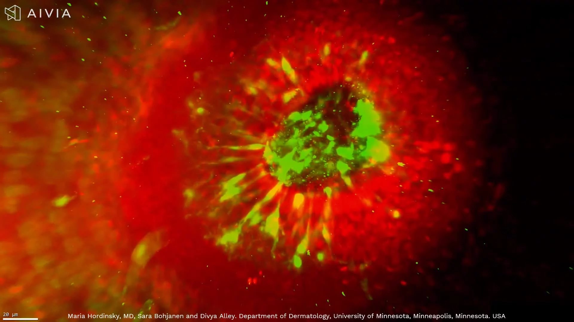
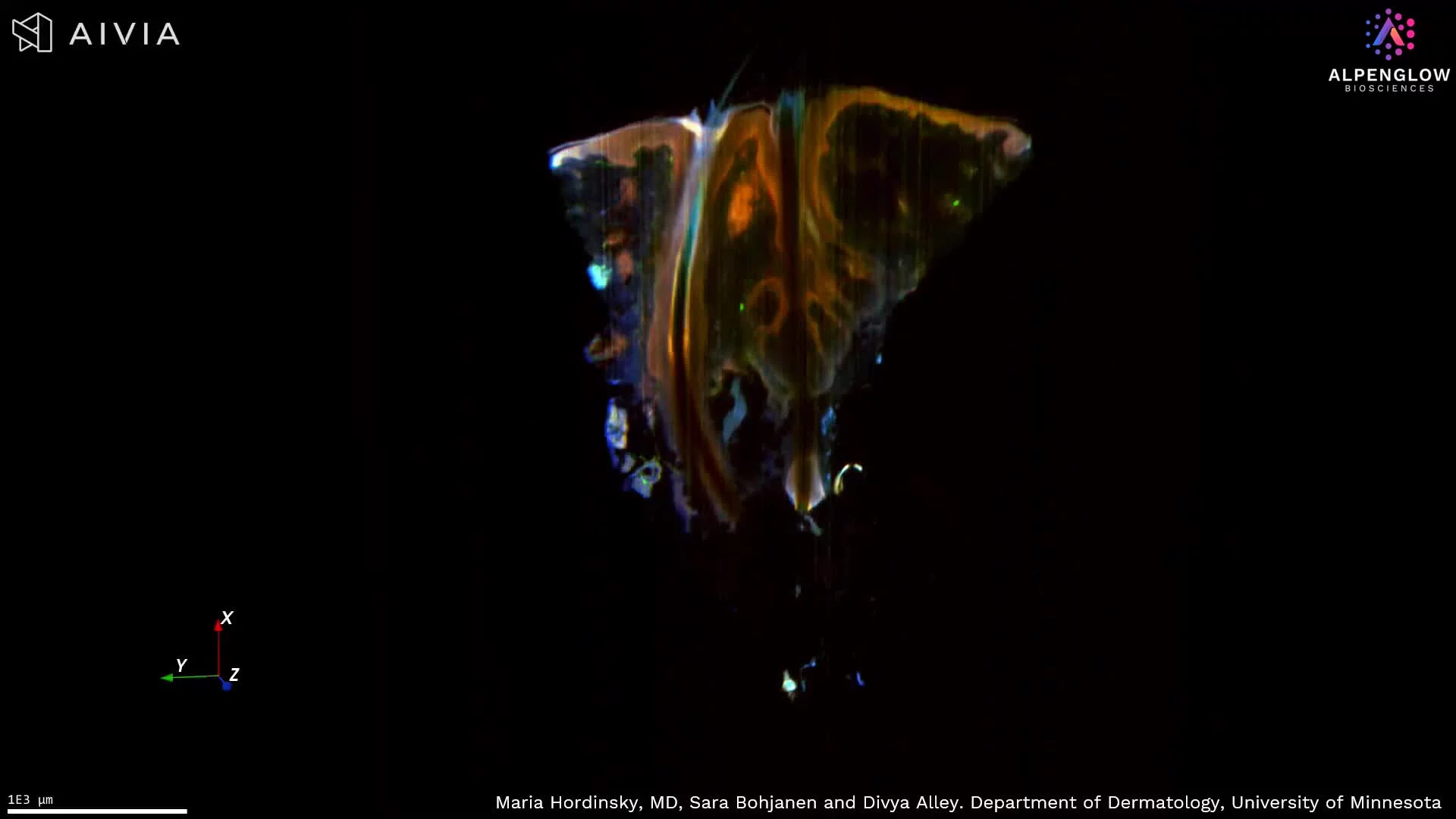
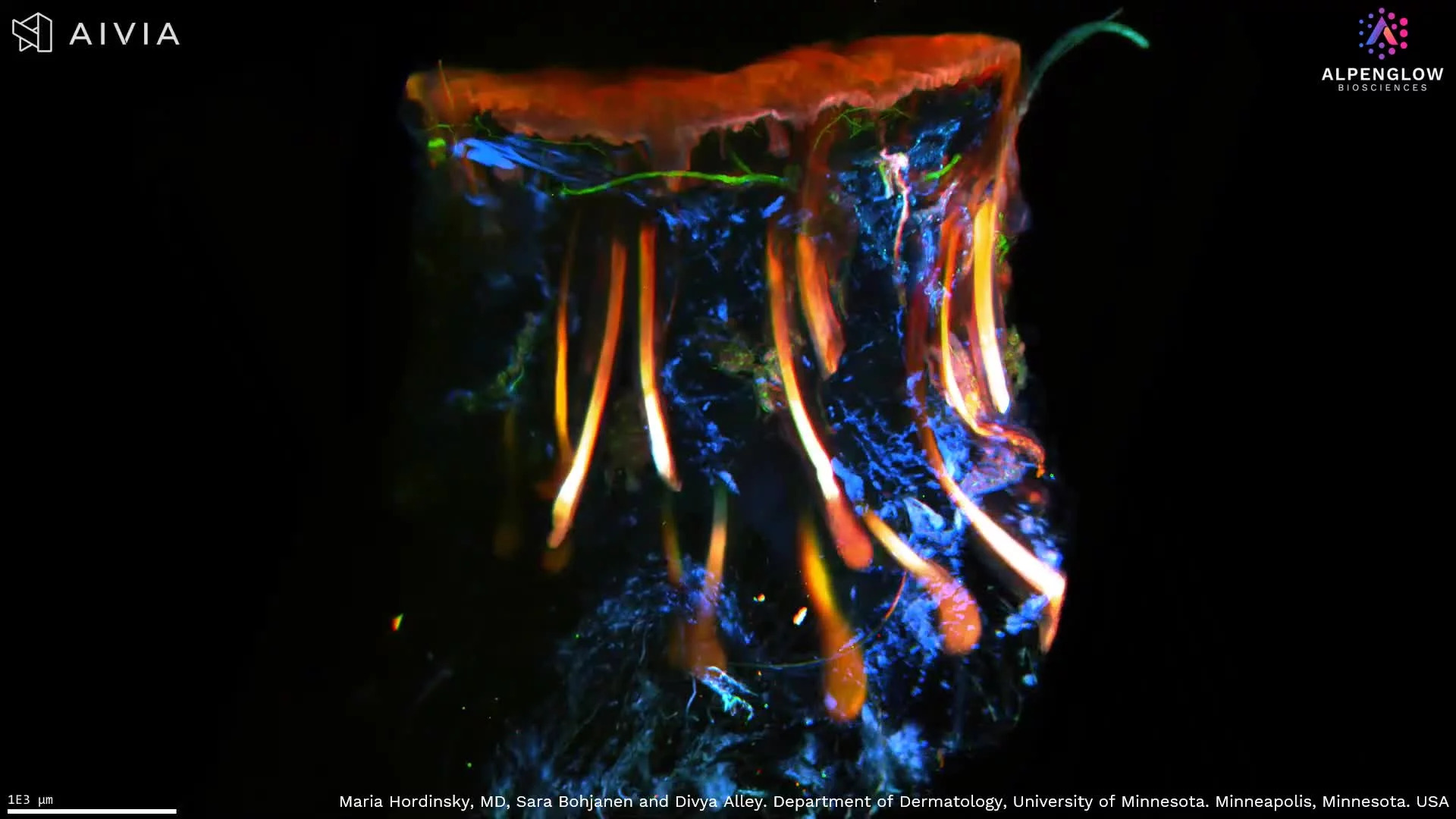
This high-resolution 3D visualization of lesional atopic dermatitis skin tissue reveals the spatial relationship between cutaneous nerves and CD45-positive immune cells.
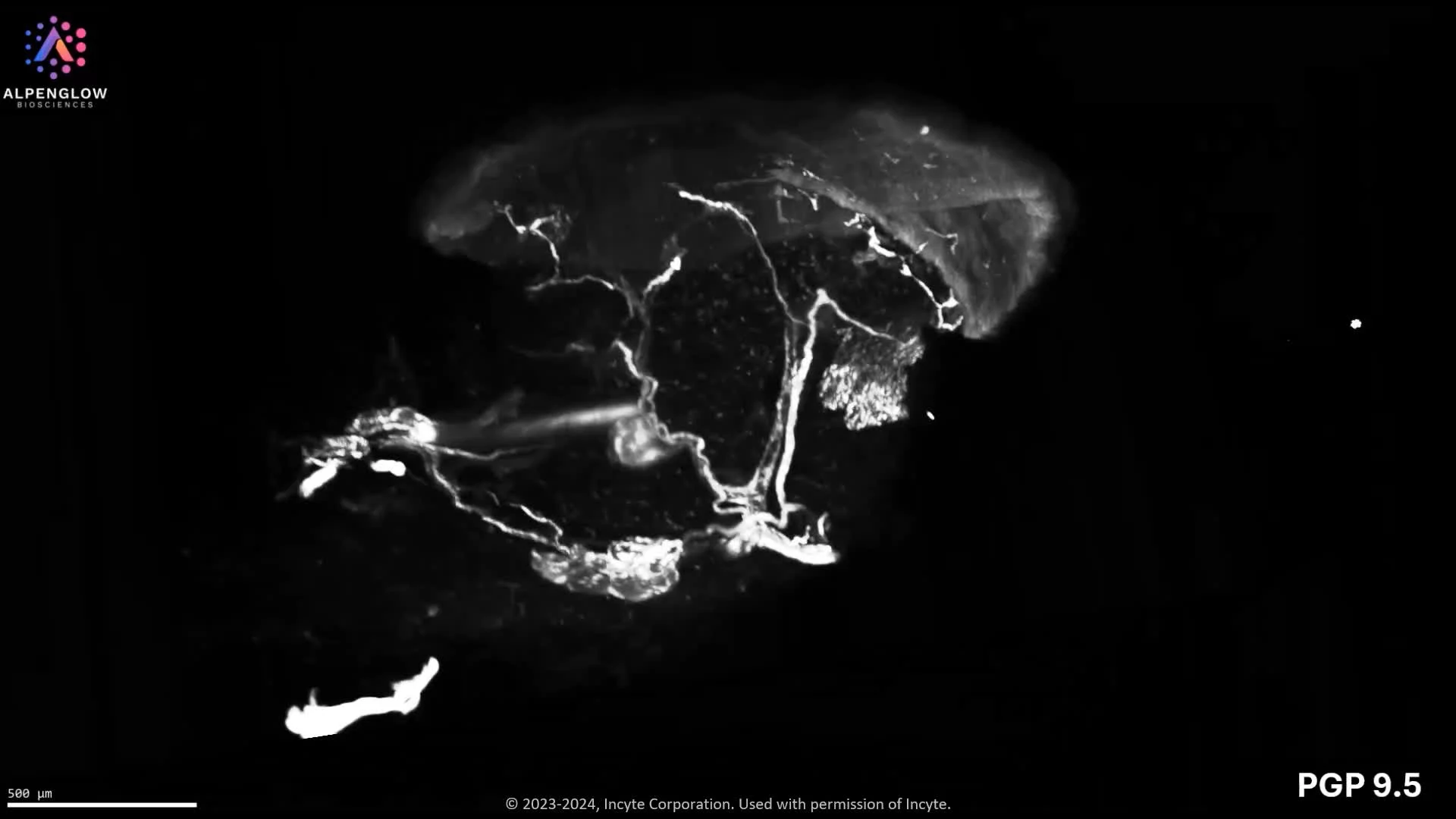
Using high-resolution 40X HOTLS imaging, TO-PRO-3, shown in blue, labels nuclei. PGP9.5, shown in white, traces nerve fibers, while CD45, shown in yellow, highlights immune cells across the tissue volume.
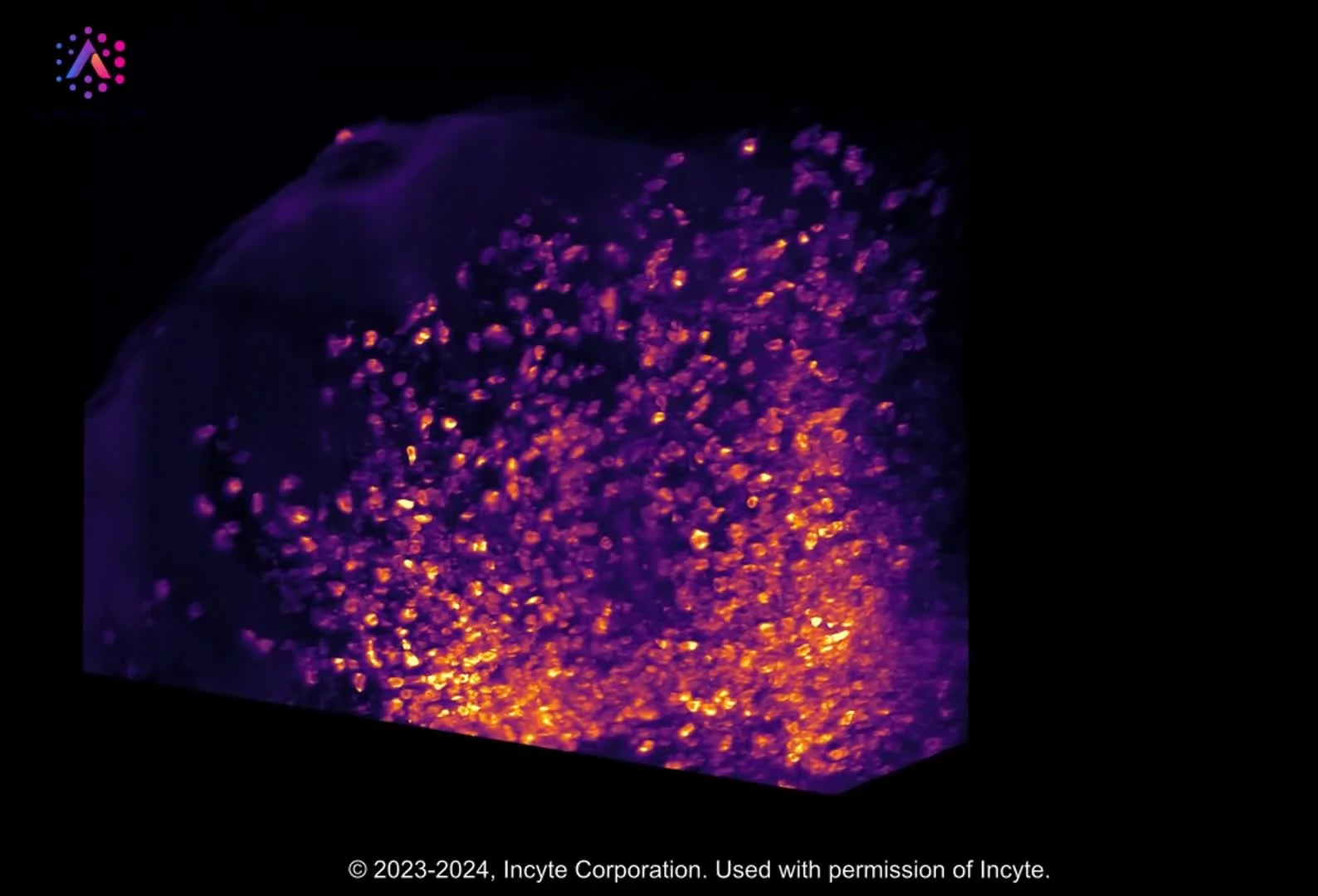
The volumetric view preserves epidermal and dermal architecture while revealing clusters of CD45-positive immune cells near selected nerve fibers across tissue depth. This spatial proximity provides a tissue-wide view of neuro-immune organization without implying a functional interaction from imaging alone.
With appropriate segmentation, the dataset supports measurement of immune-cell density, nerve density, clustering, distances between immune cells and nerves, and regional variation across skin compartments. These readouts are relevant to research on inflammatory skin diseases and the neuroimmune mechanisms underlying itch and inflammation.
Explore how 3D tissue imaging supports dermatology research across skin architecture, innervation, immune organization, and spatial relationships.
The tissue was imaged using the Aurora 3D™ Spatial Biology Solution, including the 3Di™ Hybrid Open-Top Light-Sheet microscope.