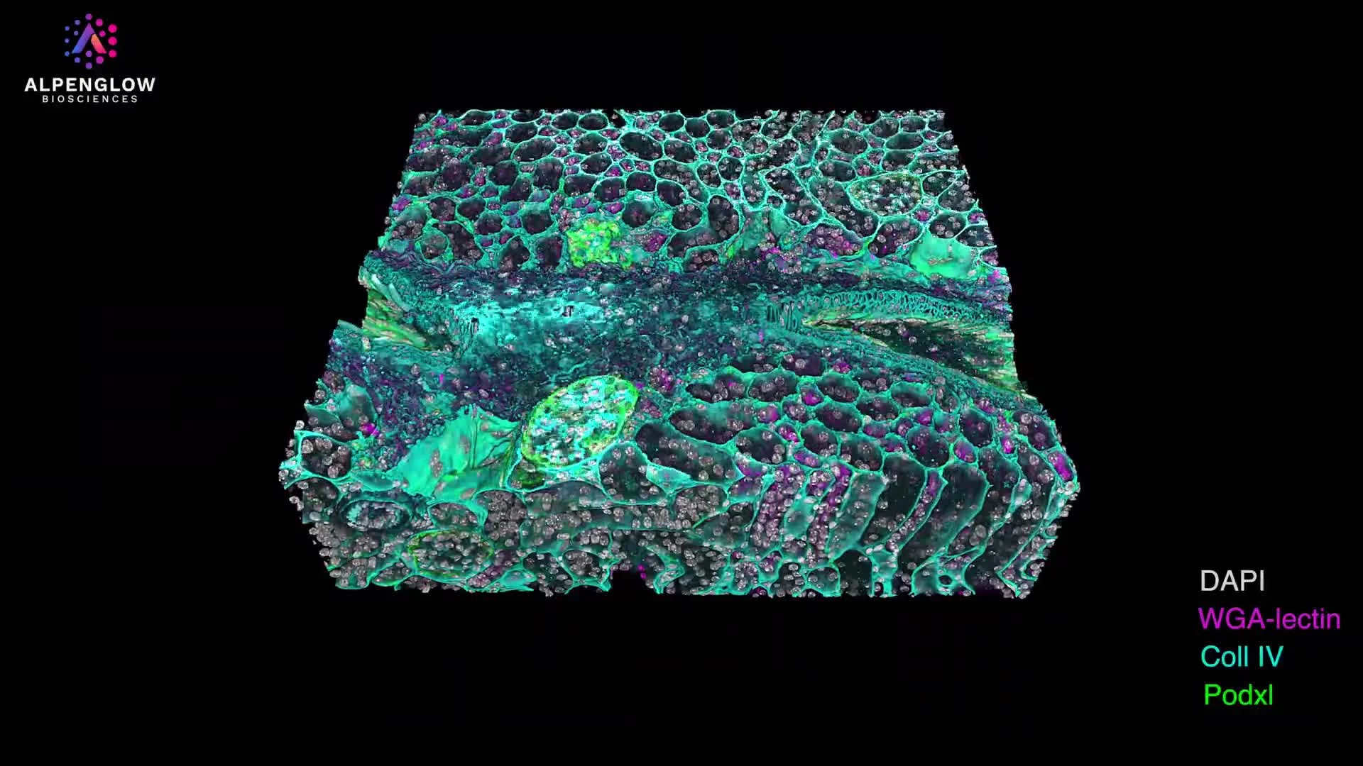
3D Imaging of Vascular and Mural Cell Architecture in a Whole Murine Heart
This dataset presents a whole murine heart imaged in 3D, preserving vascular and vessel-associated cellular architecture across the intact organ.
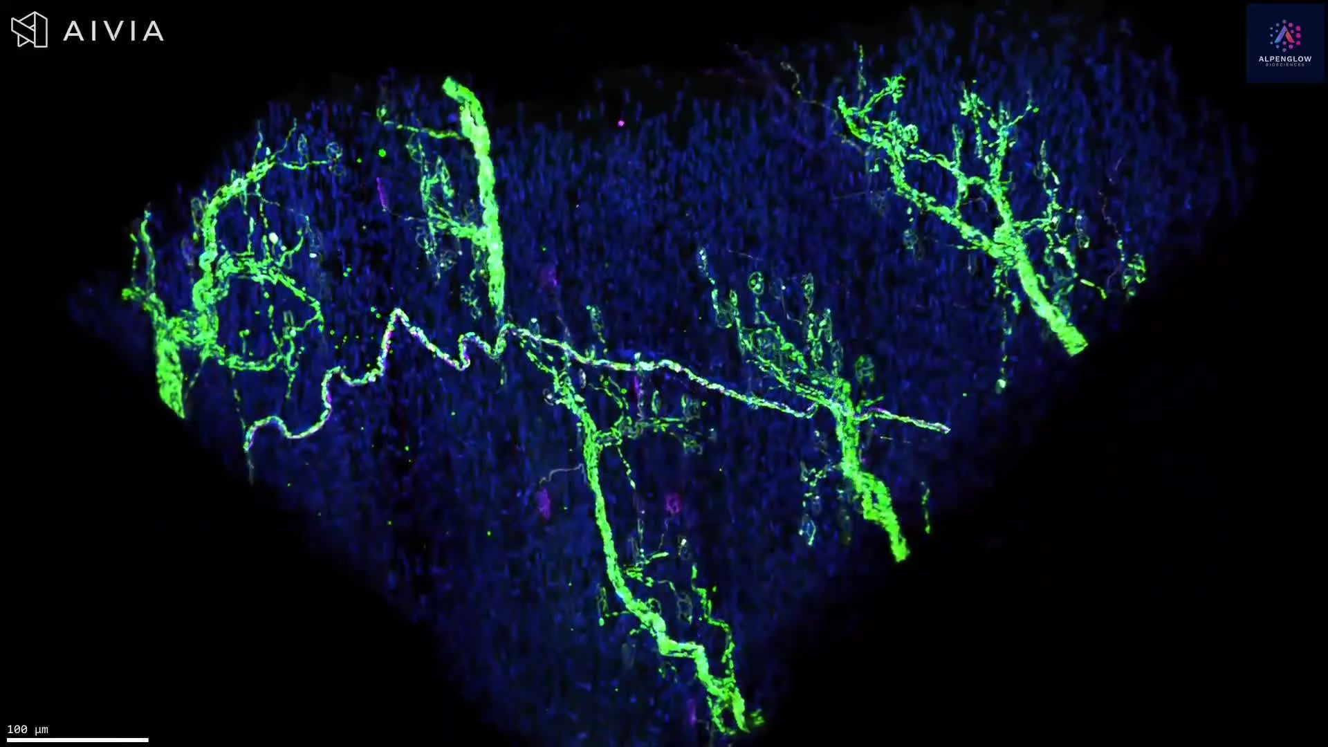
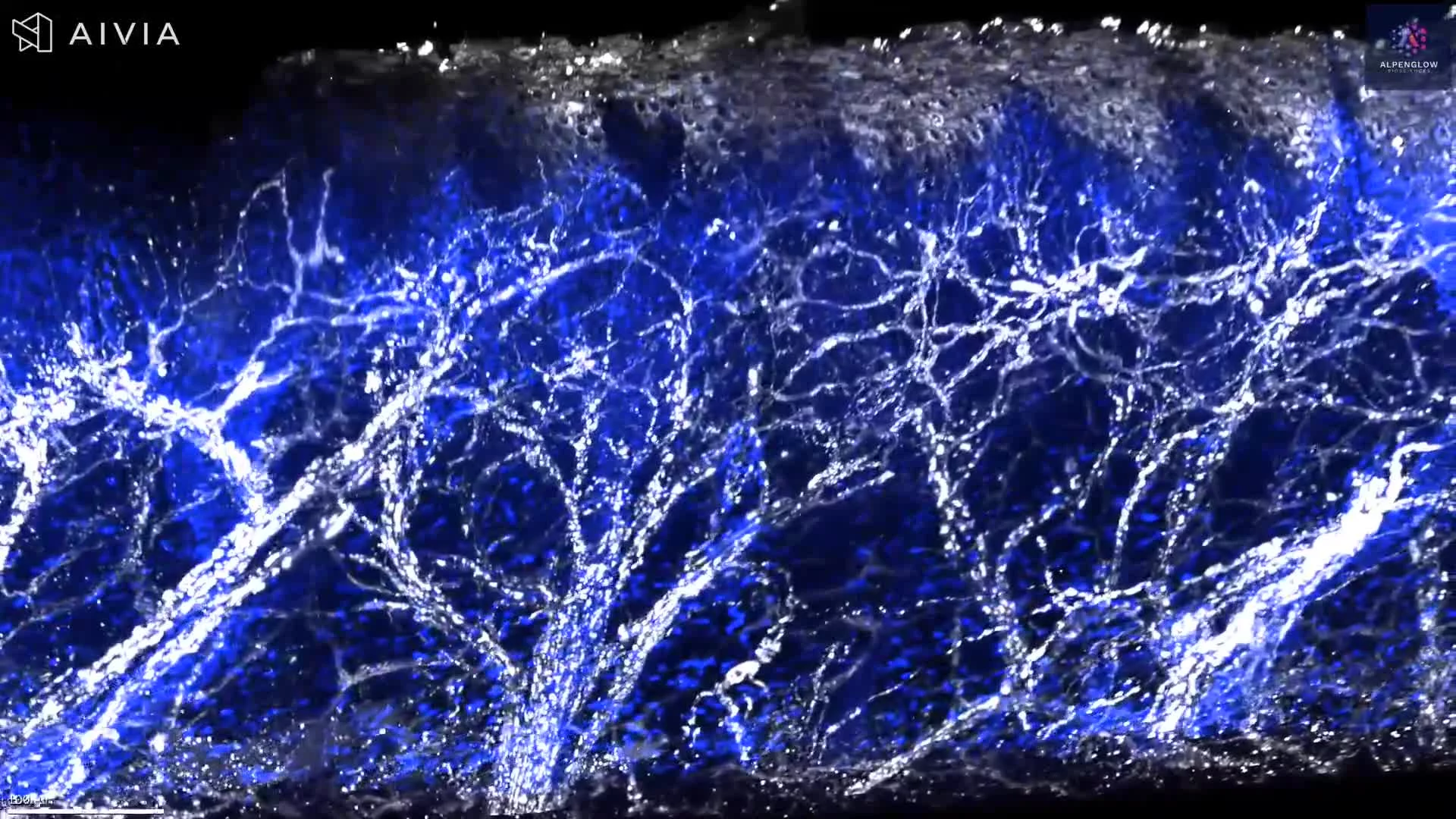
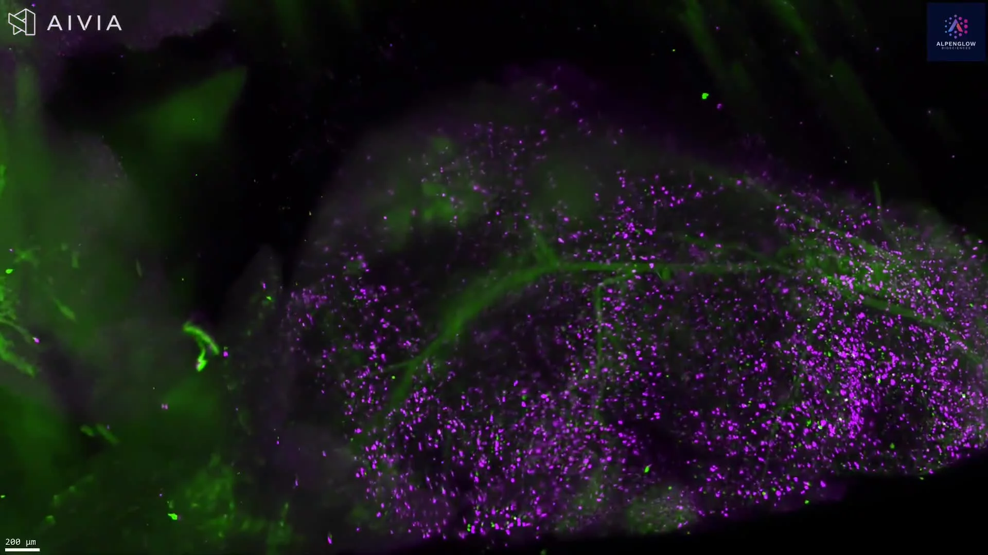
Smooth muscle actin, shown in green, highlights SMA-positive structures associated with the vasculature. A mural-cell-associated signal, shown in purple, reveals vessel-associated cells along the vascular network.
The video begins with larger vessels and progressively moves into selected regions, combining Scout imaging of the whole organ with high-resolution Zoom imaging of local vascular structures. This multiscale view reveals vessel branching and the organization of mural cells around vessels, including fine perivascular structures at cellular detail.
The dataset supports quantitative analysis of vessel density, branching, diameter, tortuosity, mural-cell distribution, and spatial proximity between vessel-associated cells and the vascular wall. These measurements are relevant to cardiovascular research, vascular biology, tissue remodeling, and preclinical studies of disease and treatment response.
The tissue was imaged using the Aurora 3D™ Spatial Biology Solution, including the 3Di™ Hybrid Open-Top Light-Sheet microscope.