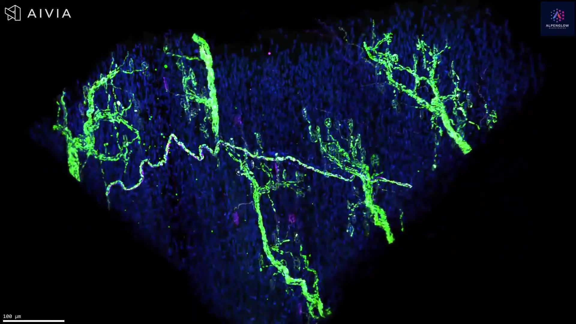
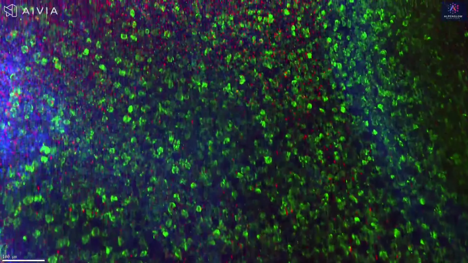
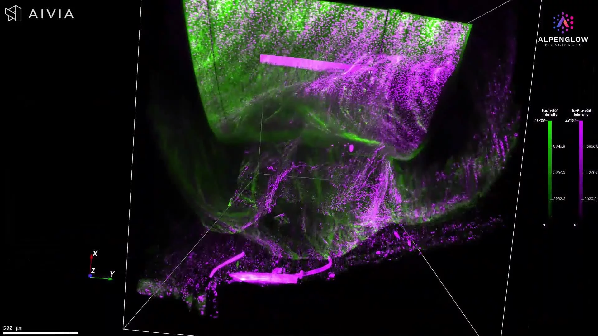
3D Imaging of Pig Muscle Section Stained with CD31 and YO-PRO-1
This video showcases a 2 mm section of pig muscle captured in 3D using the Aurora™ 3Di Hybrid Open Top Light Sheet (HOTLS) microscope. The tissue was stained to visualize endothelial and nuclear structures, enabling volumetric mapping of the vascular system.
Stains used:
CD31 (Green): Marks endothelial cells and vasculature
YO-PRO-1 (Red): Labels cell nuclei
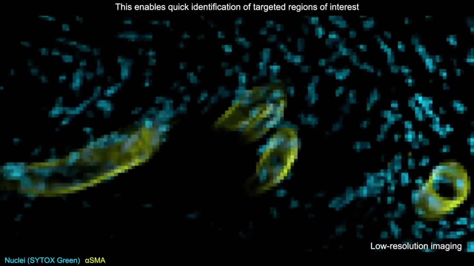
The dataset demonstrates the flexibility of the Scout + Zoom workflow, transitioning from rapid low-resolution scans for whole-sample context to detailed high-resolution imaging of selected regions. This approach reveals the intricate vascular network in full three-dimensional context, exposing insights that conventional 2D histology cannot provide.
By combining 3D histology with 3Dm data management and 3Dai AI-powered segmentation, researchers can quantify vessel density, branching, and spatial relationships with precision. Applications include preclinical vascular biology, muscle physiology, and translational research where accurate vascular mapping is critical.