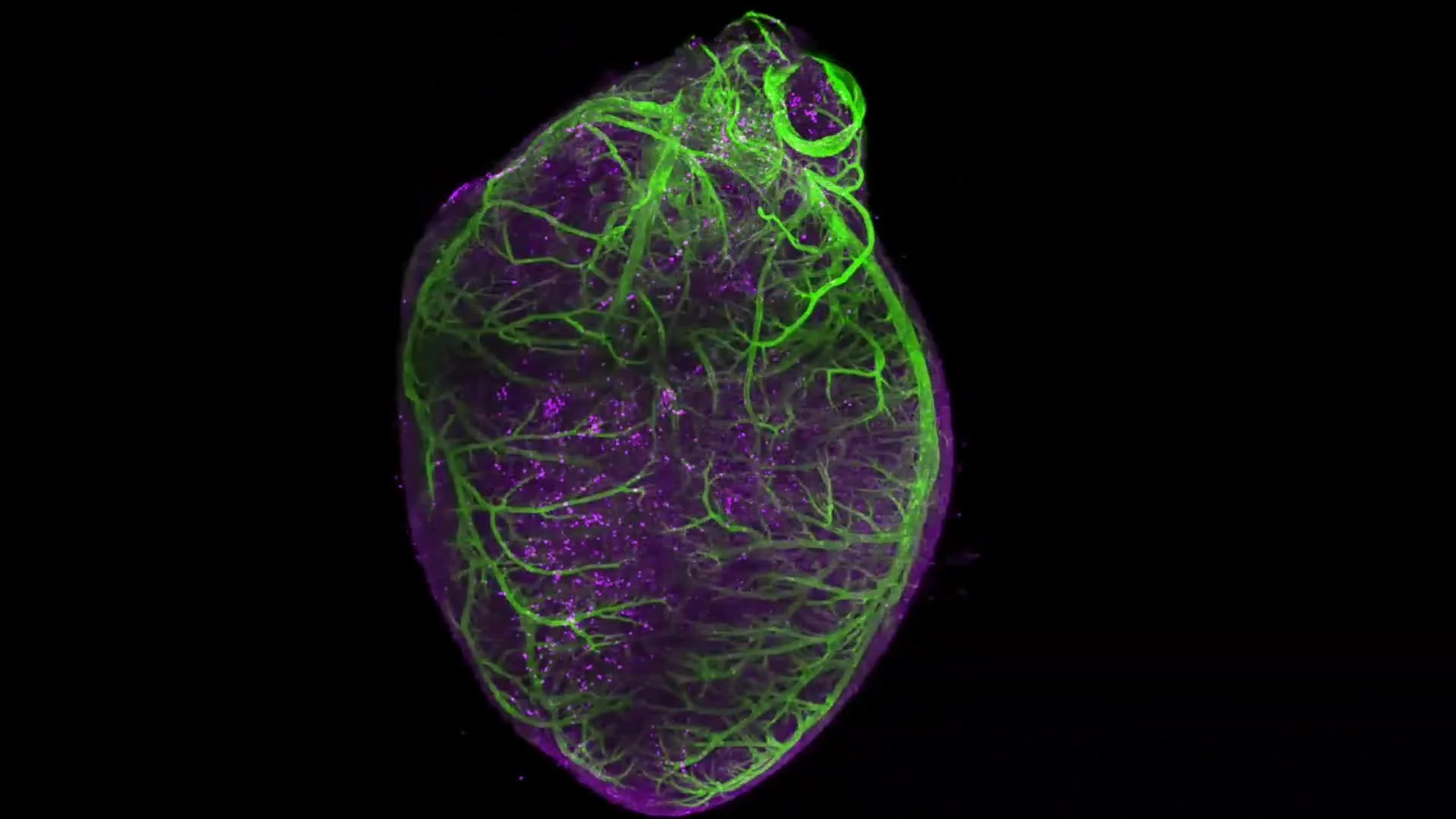
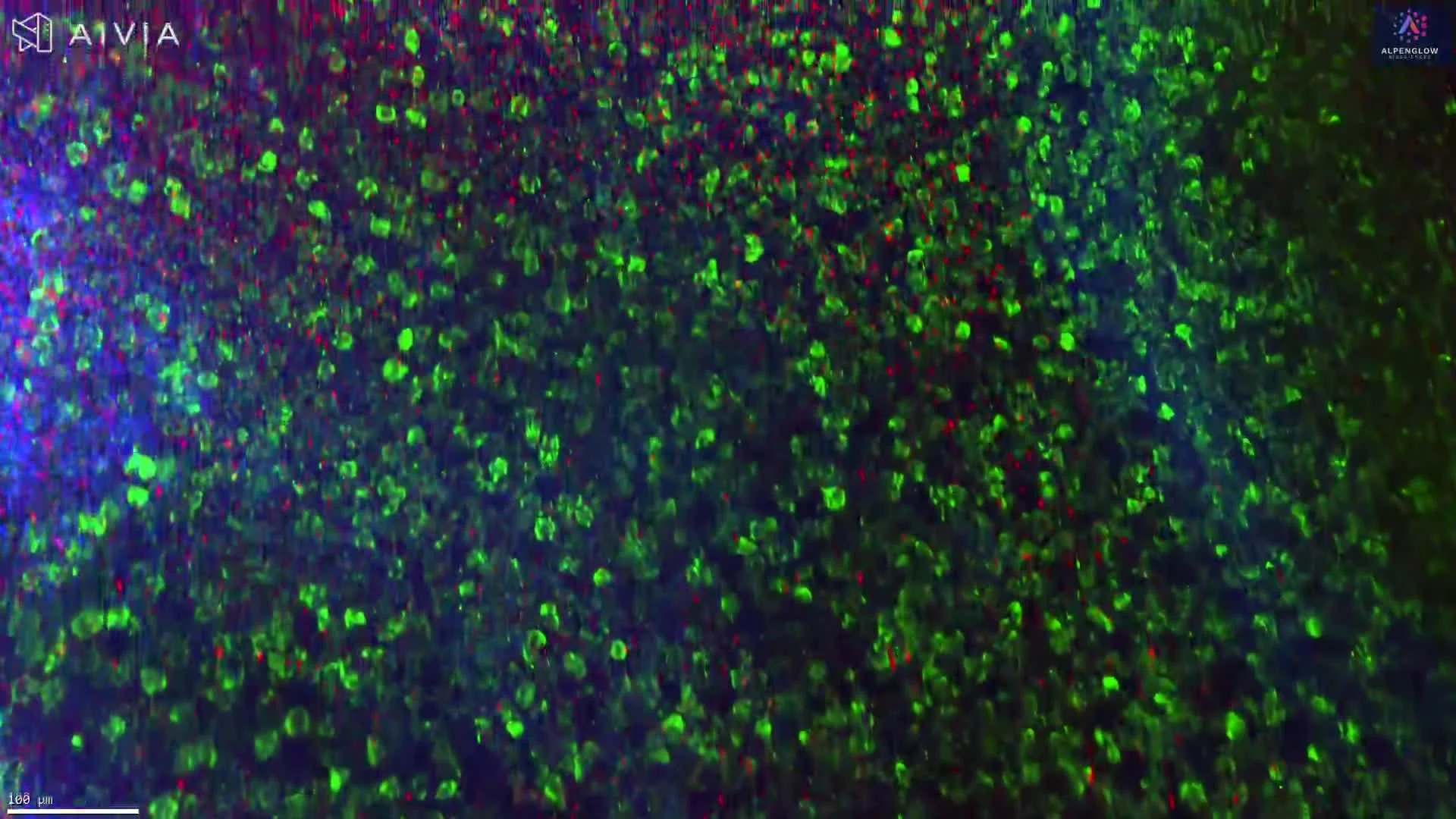
3D Imaging of Mouse Pancreatic Cyst with Ki67 and γ-H2AX
This high-resolution 3D dataset showcases a mouse pancreatic cyst dual-stained to reveal key biological processes:
Stains used:
Ki67 (Green): Highlights proliferating cells
γ-H2AX (Purple): Marks sites of DNA damage
Captured with the Aurora™ 3Di Hybrid Open Top Light Sheet (HOTLS) microscope, the dataset preserves intact tissue structure, enabling visualization of the cyst architecture in full volumetric detail. Unlike conventional 2D histology, this 3D workflow eliminates sampling bias and retains spatial relationships essential for accurate biological interpretation.
Visualize the complete cyst structure without missing features due to sectioning
Accurately localize proliferating and damaged cells in their native context
Maintain spatial relationships often lost in thin slices
Combined with 3Dm data management and 3Dai AI-powered segmentation, this workflow delivers quantifiable insights into proliferation, DNA damage, and microenvironmental context.
Applications include cancer research, pancreatic disease modeling, and translational studies where accurate mapping of cystic structures is critical.