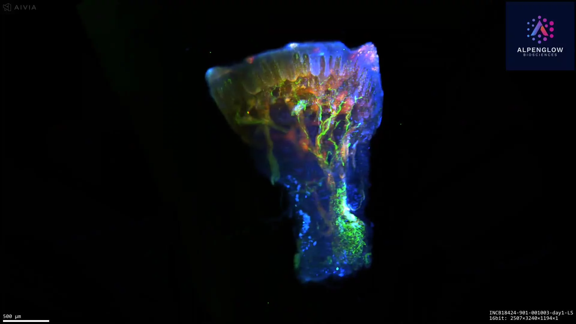
3D imaging of nerve innervation at the dermal–epidermal junction
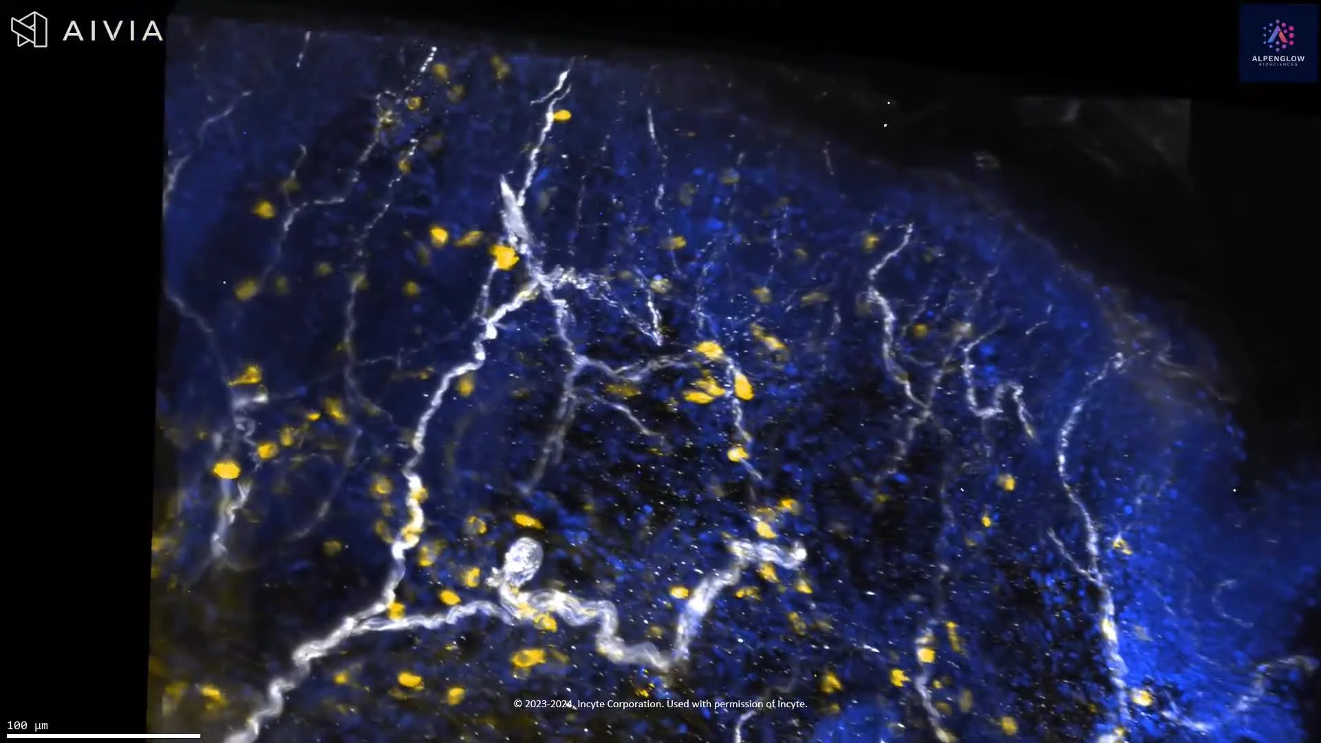
This dataset presents volumetric 3D imaging of rat toe pad skin, revealing cutaneous nerve architecture at the dermal–epidermal junction.
PGP9.5 highlights nerve fibers in white, while YO-PRO-1 labels nuclei in blue. The intact tissue volume preserves the continuity of larger nerve bundles, finer branches, fiber crossings, and terminal structures across tissue depth.
With appropriate segmentation, the dataset can support quantitative analysis of nerve density, fiber length, branching, orientation, and the distribution of nerve fibers relative to the epidermis and surrounding cells. These measurements are relevant to studies of cutaneous innervation, sensory biology, neuro-immune interactions, pain, itch, and disease-associated changes in nerve architecture.
The tissue was imaged using the Aurora 3D™ Spatial Biology Solution, including the 3Di™ Hybrid Open-Top Light-Sheet microscope.