3D Imaging of Human Duodenum Villi
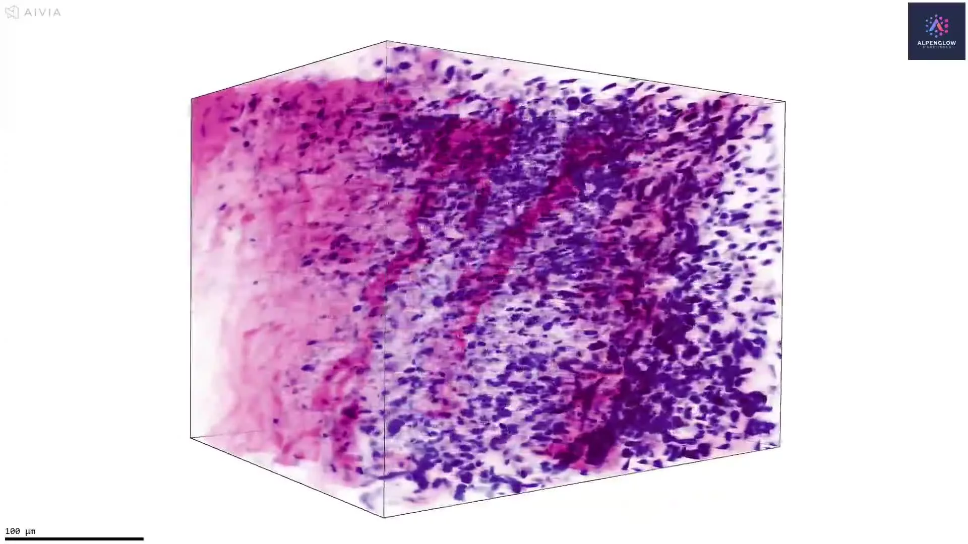
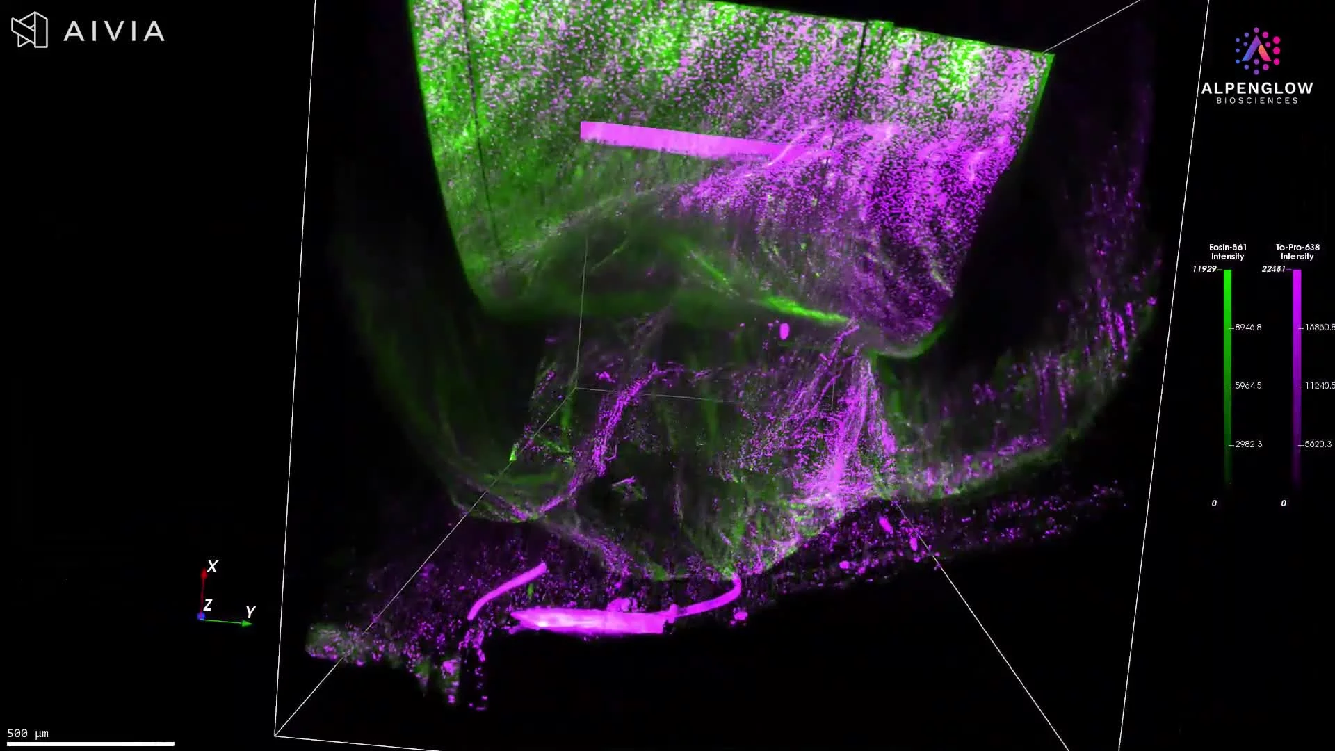
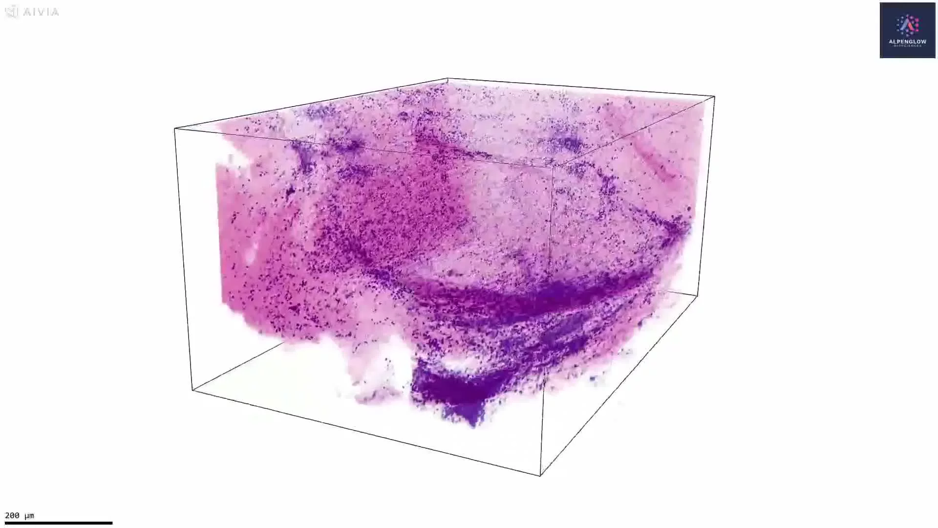
This video presents a volumetric visualization of human duodenum tissue stained with eosin and the nuclear marker TO-PRO-3, then pseudocolored to create an H&E-like appearance.
3D tissue imaging reveals intestinal villi as continuous structures across the imaged volume, preserving information about their morphology, orientation, spacing, density, and regional organization. Viewing villi across depth also reduces the influence of sectioning angle and sampling location that can affect measurements from individual 2D sections.
With appropriate segmentation, the dataset can support quantitative analysis of villus number, height, width, volume, elongation, spacing, density, and variation across tissue regions.
These measurements are relevant to gastrointestinal research, including studies of celiac disease, where villous architecture may become shortened, flattened, fused, or otherwise disrupted. Quantifying these changes across a tissue volume could help characterize the distribution and heterogeneity of structural alterations.
The tissue was imaged on the Aurora 3D™ platform using the 3Di™ Hybrid Open-Top Light-Sheet (HOTLS) microscope.