

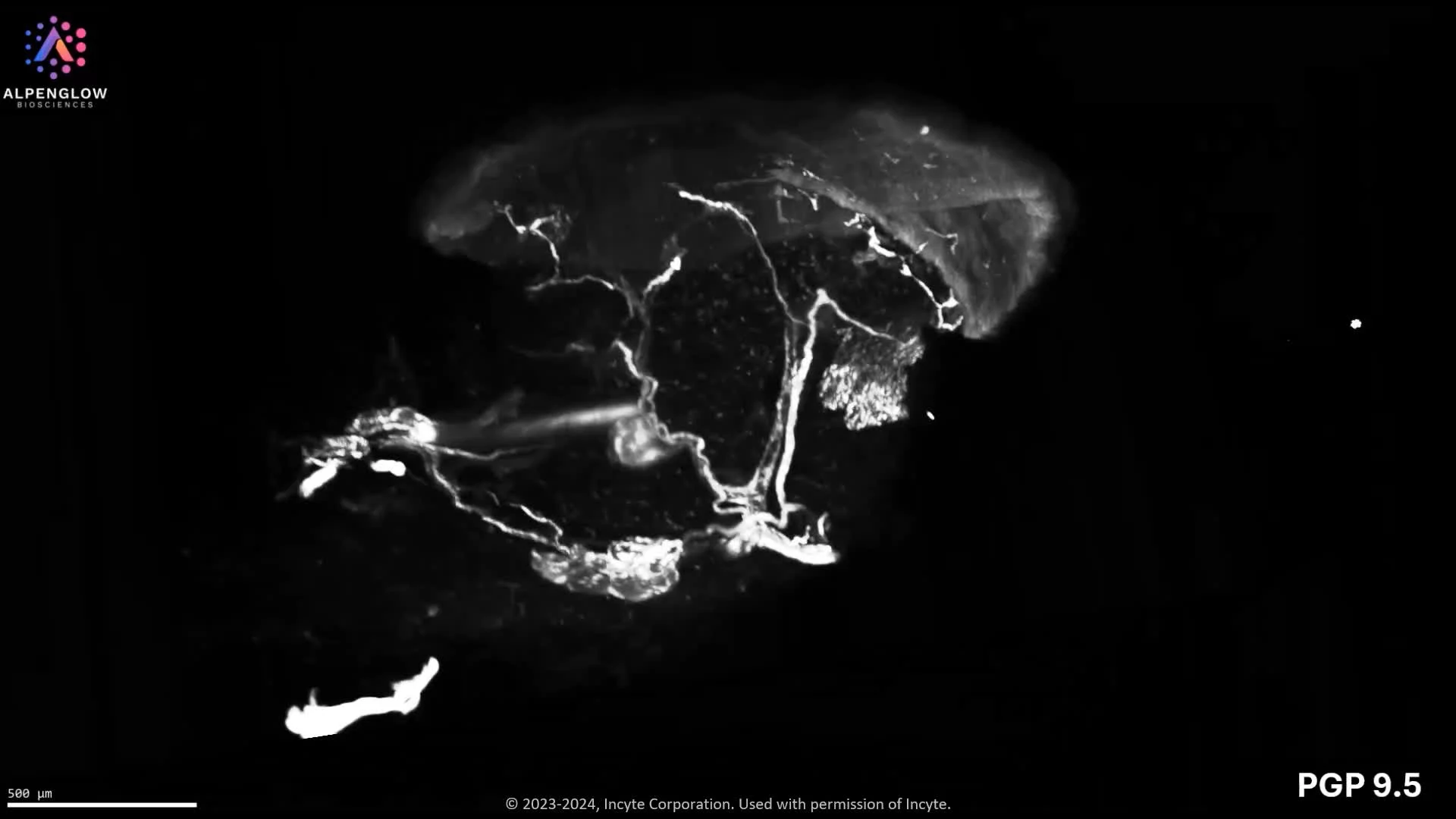
3D fluorescence imaging of mouse skin reveals complex nerve branching across intact tissue, enabling quantitative spatial analysis beyond 2D histology.
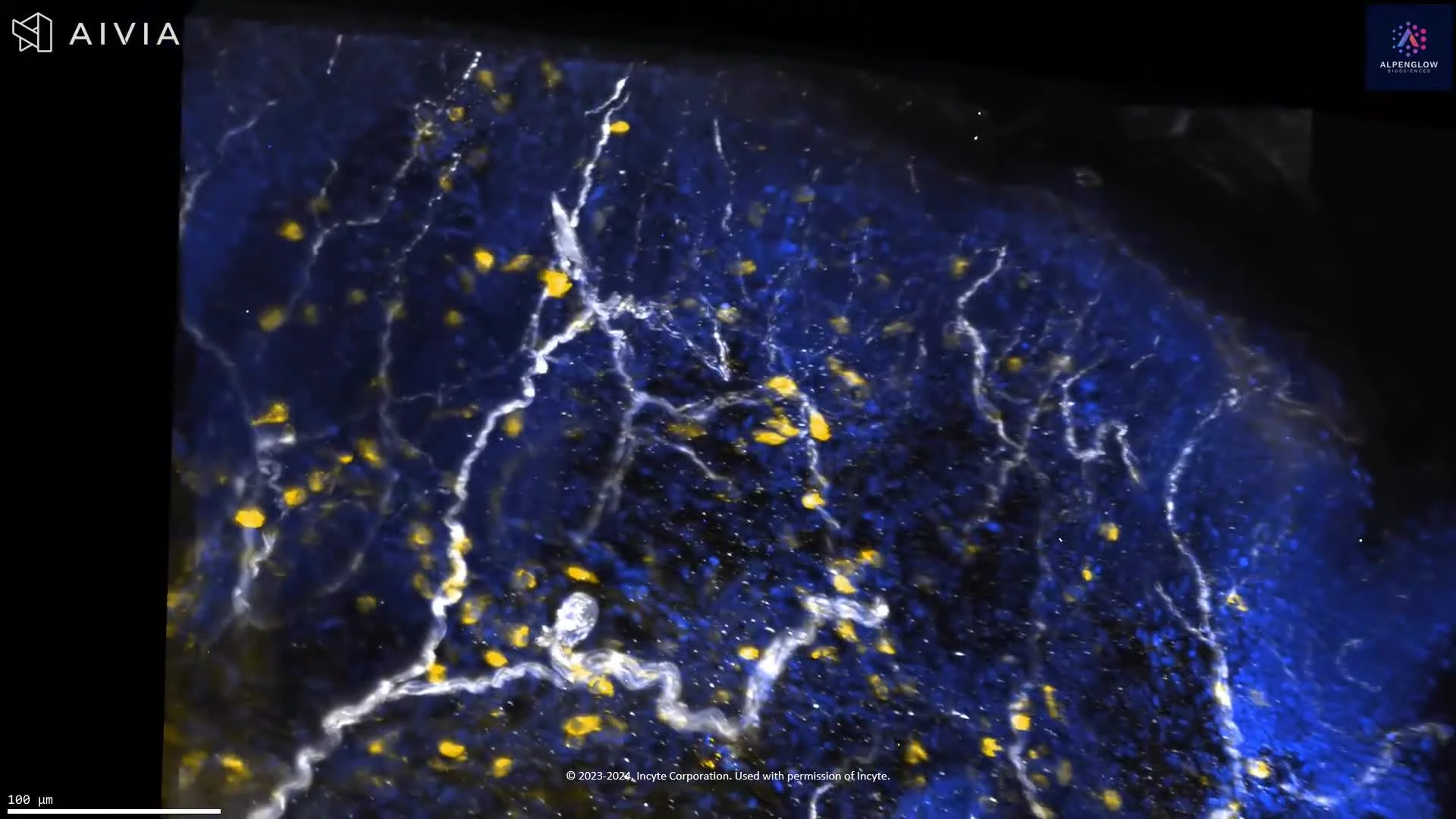
High-resolution 3D imaging of nerve networks at the dermal–epidermal junction reveals complex branching and spatial organization across intact skin tissue.
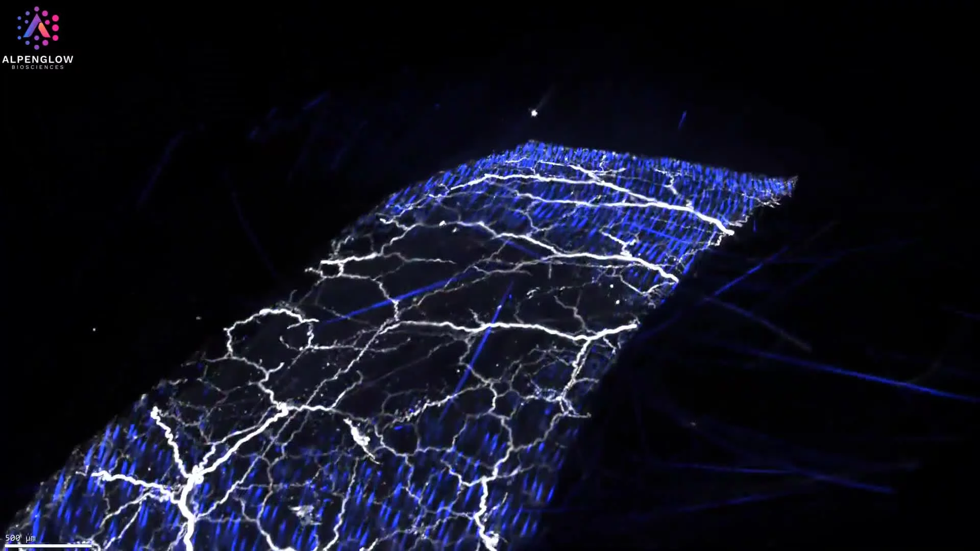
This 3D video reveals the intricate branching architecture of peripheral nerves in mouse skin using Yo-Pro-1 and PGP9.5 staining, highlighting how 3D intact tissue imaging captures complex neural networks across the full tissue depth.
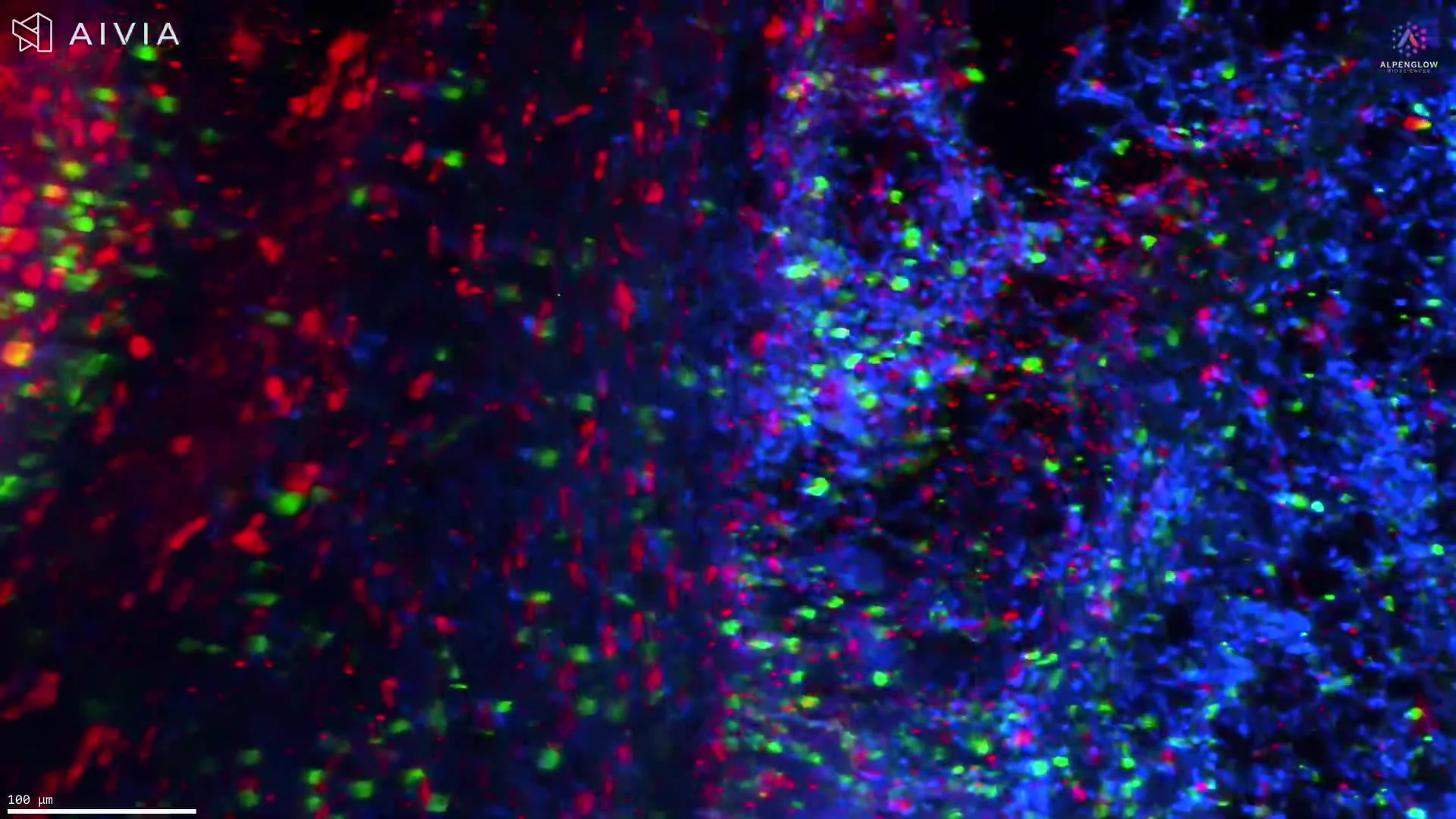
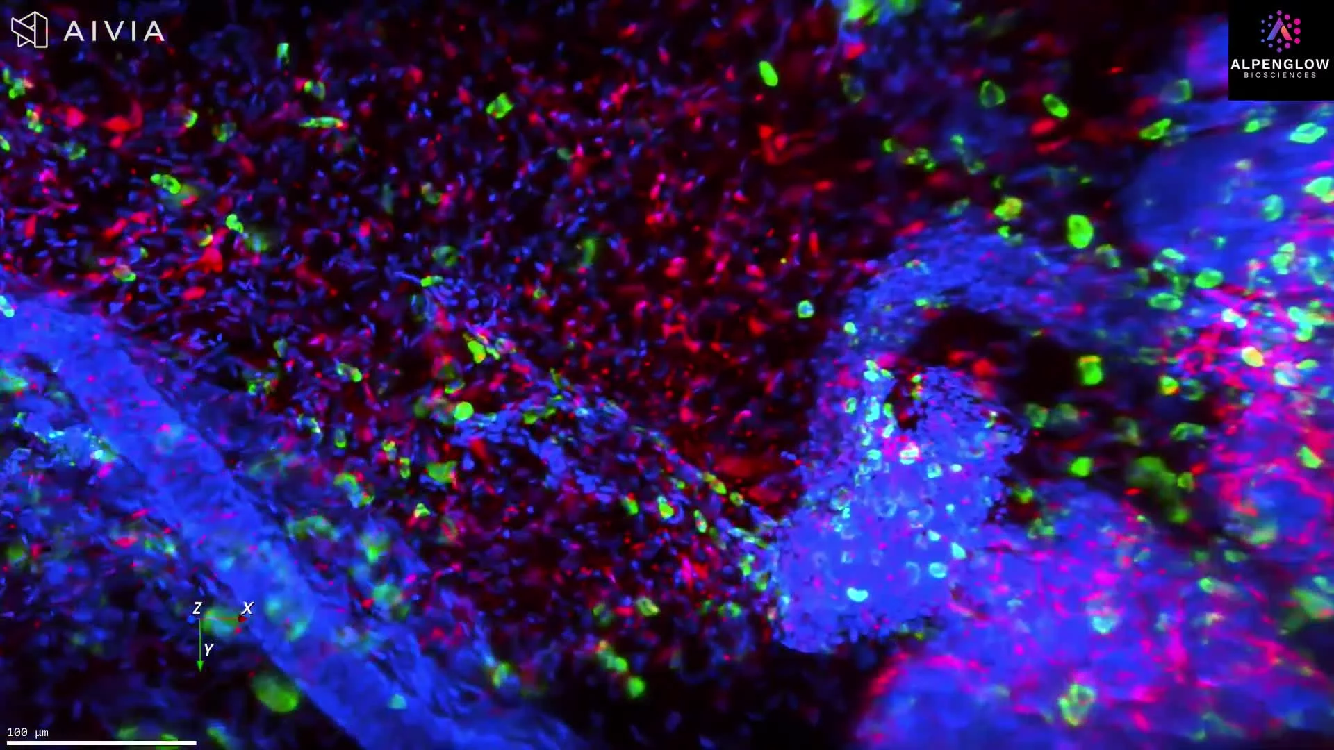
High-resolution 3D imaging of CD45-stained skin biopsy shows lymphocyte clustering around nerves, offering new insights into immune cell behavior.
High-resolution 3D imaging of scalp tissue reveals nuclei, immune cells, and nerve networks, driving new insights into alopecia and other skin diseases.
High-resolution 3D imaging of atopic dermatitis reveals lymphocyte clusters near nerves, stained with TO-PRO-3, PGP 9.5, and CD45. Explore detailed innervation and immune-cell interactions in lesional skin.
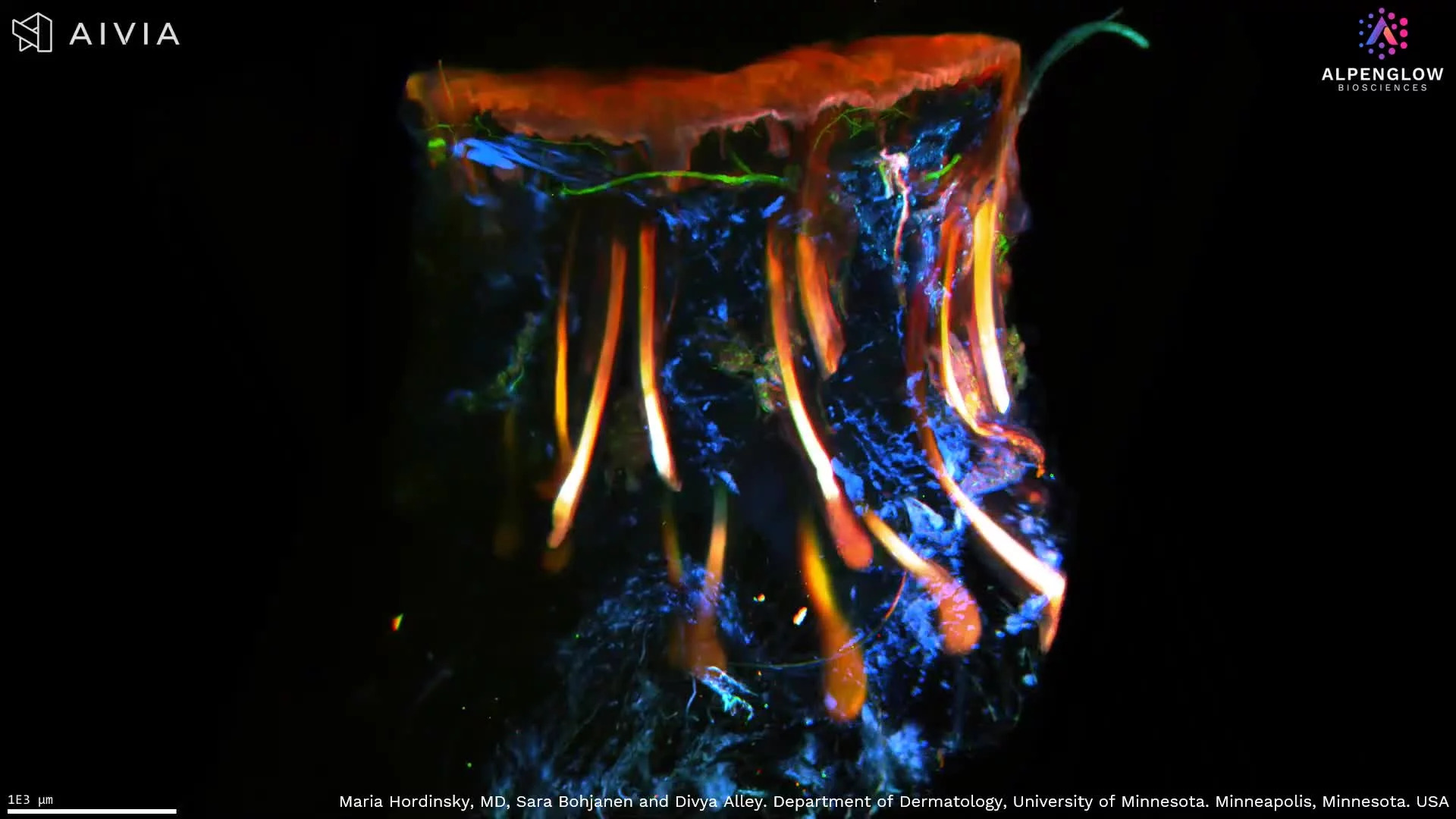
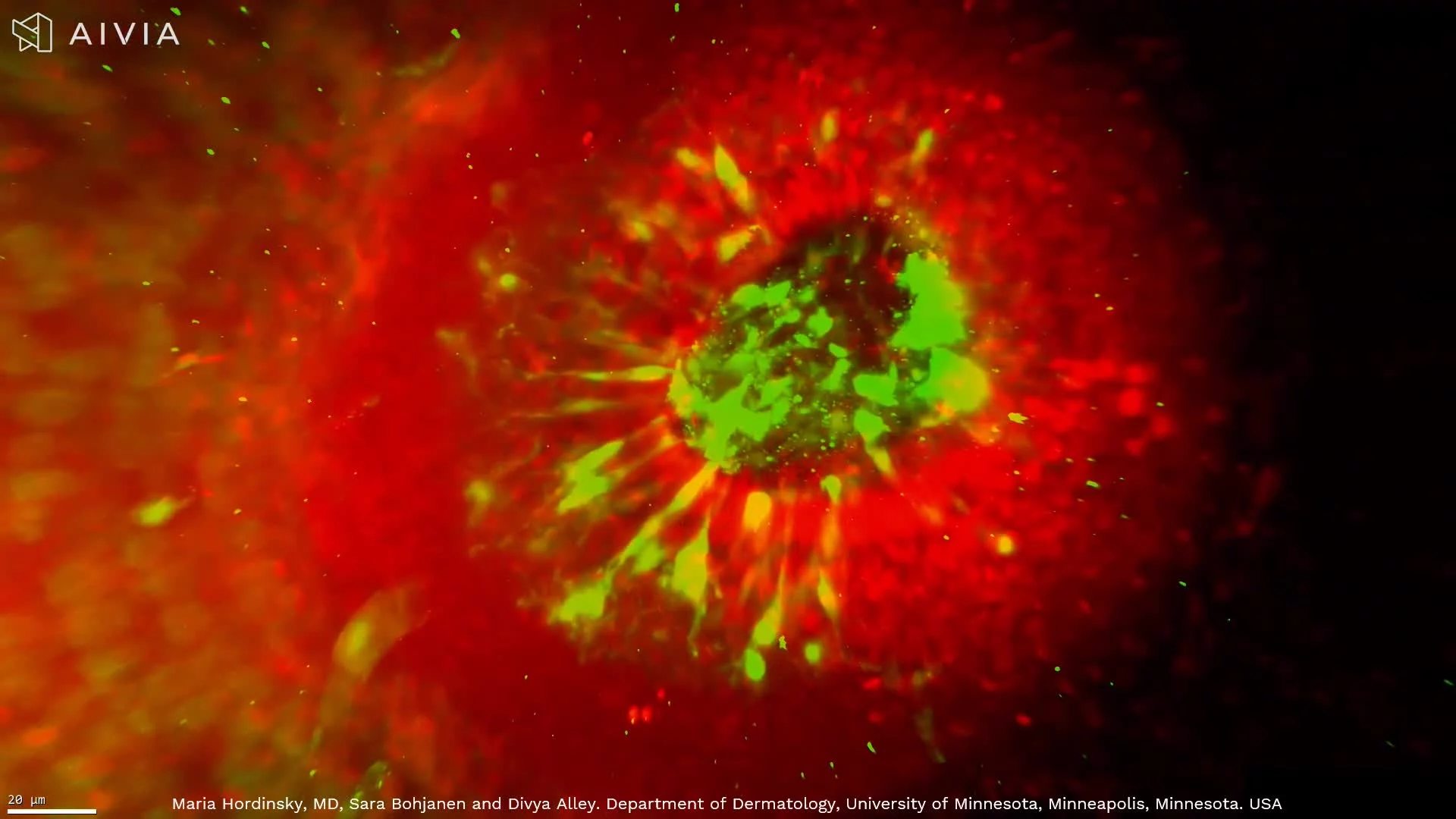
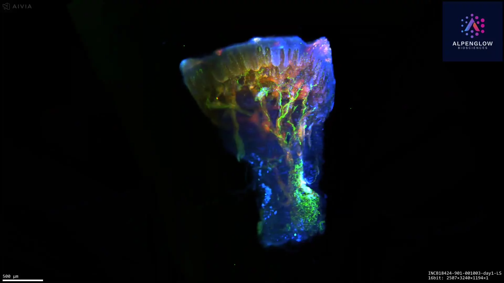
3D imaging of skin reveals a hair follicle, its gland, and nerve network with stunning clarity, advancing research in alopecia and inflammatory skin disease.
See hair follicles in true 3D, from bulb to dermal papilla, with detailed mapping of sensory nerves and nuclei for alopecia and hair research.High-resolution 3D imaging of scalp tissue reveals nuclei, immune cells, and nerve networks, driving new insights into alopecia and other skin diseases.
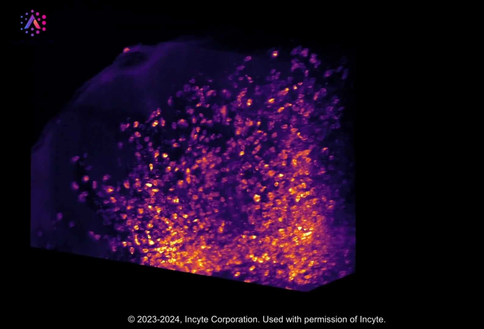
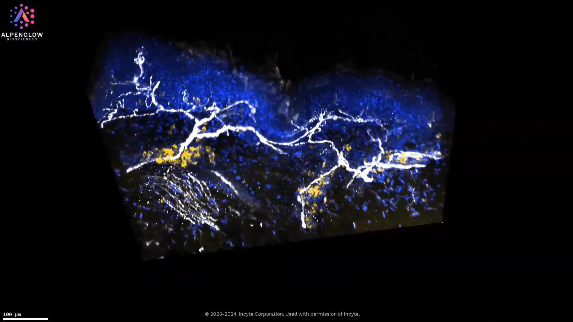
3D imaging of melanoma tissue with CD45, Neutrophil Elastase, and TO-PRO-3 reveals hidden immune–tumor interactions and sets a new standard for research.
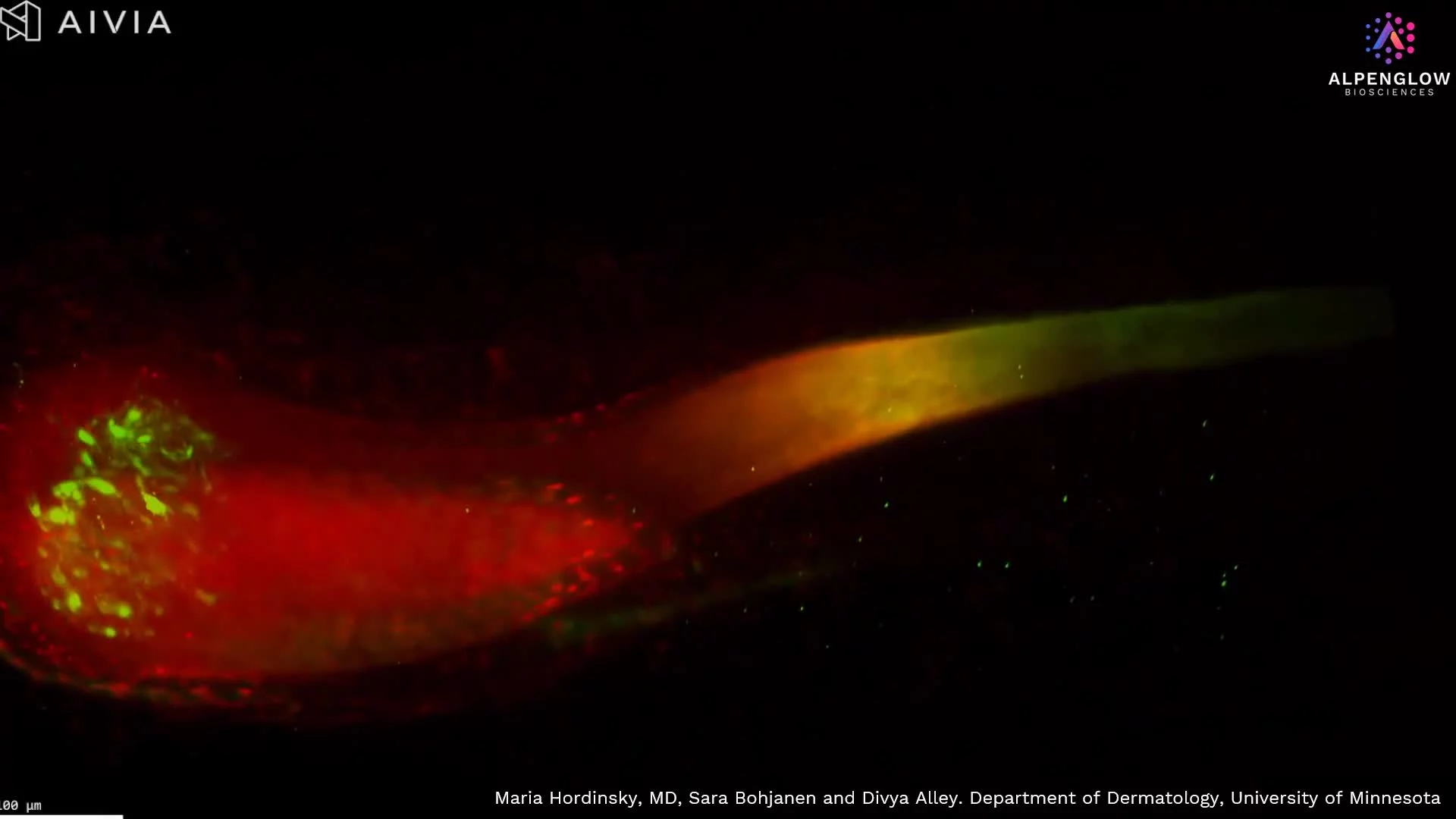
See an entire hair follicles in true 3D, from bulb to dermal papilla, with detailed mapping of sensory nerves and nuclei for alopecia and hair research.High-resolution 3D imaging of scalp tissue reveals nuclei, immune cells, and nerve networks, driving new insights into alopecia and other skin diseases.
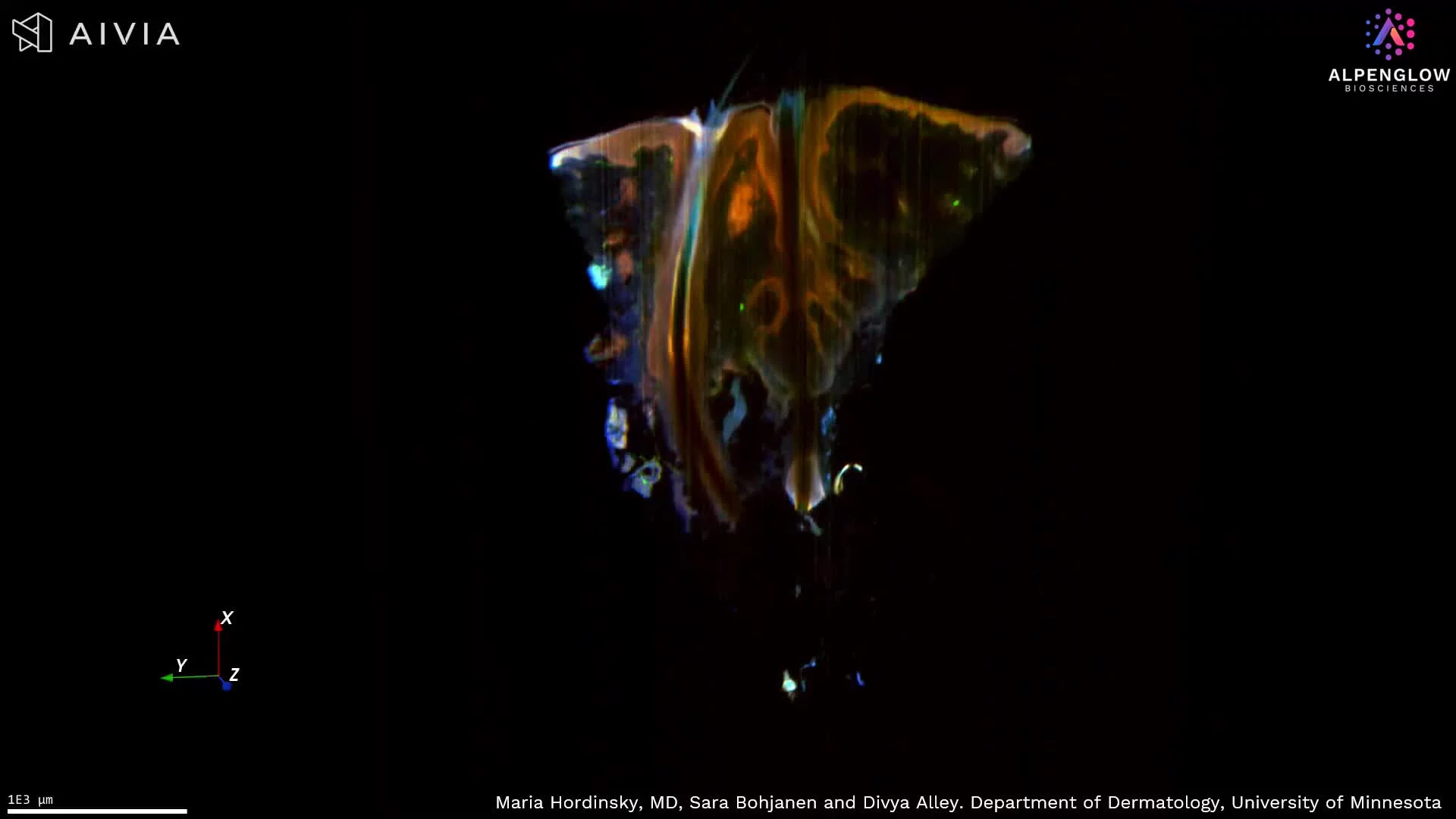
High-resolution 3D imaging of scalp tissue reveals entire follicles, nuclei, immune cells, and nerve networks, unlocking new insights into alopecia and other skin diseases.
3D imaging of atopic dermatitis skin punch biopsy; the tissue is stained with TO-PRO-3, PGP9.5, and CD45. Explore detailed innervation and immune-cell interactions in lesional skin.
3D imaging of human skin biopsy stained with tryptase, TO-PRO-3, and PGP9.5 reveals mast cell–nerve interactions for dermatology and oncology research.
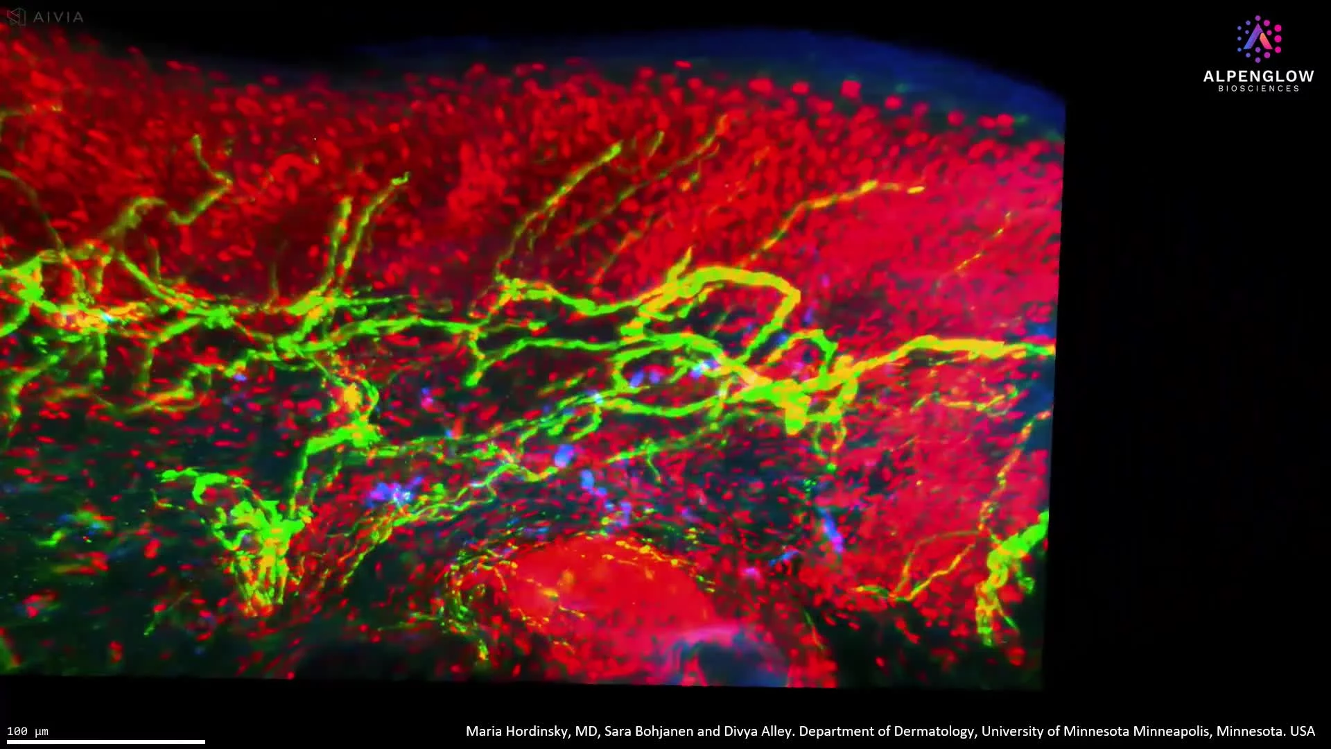
High-resolution 3D imaging of scalp epidermis reveals branching nerve structures and nuclear organization, advancing hair and dermatology research.
High-resolution 3D imaging of atopic dermatitis reveals precise innervation and immune cell distribution, surpassing conventional histology.
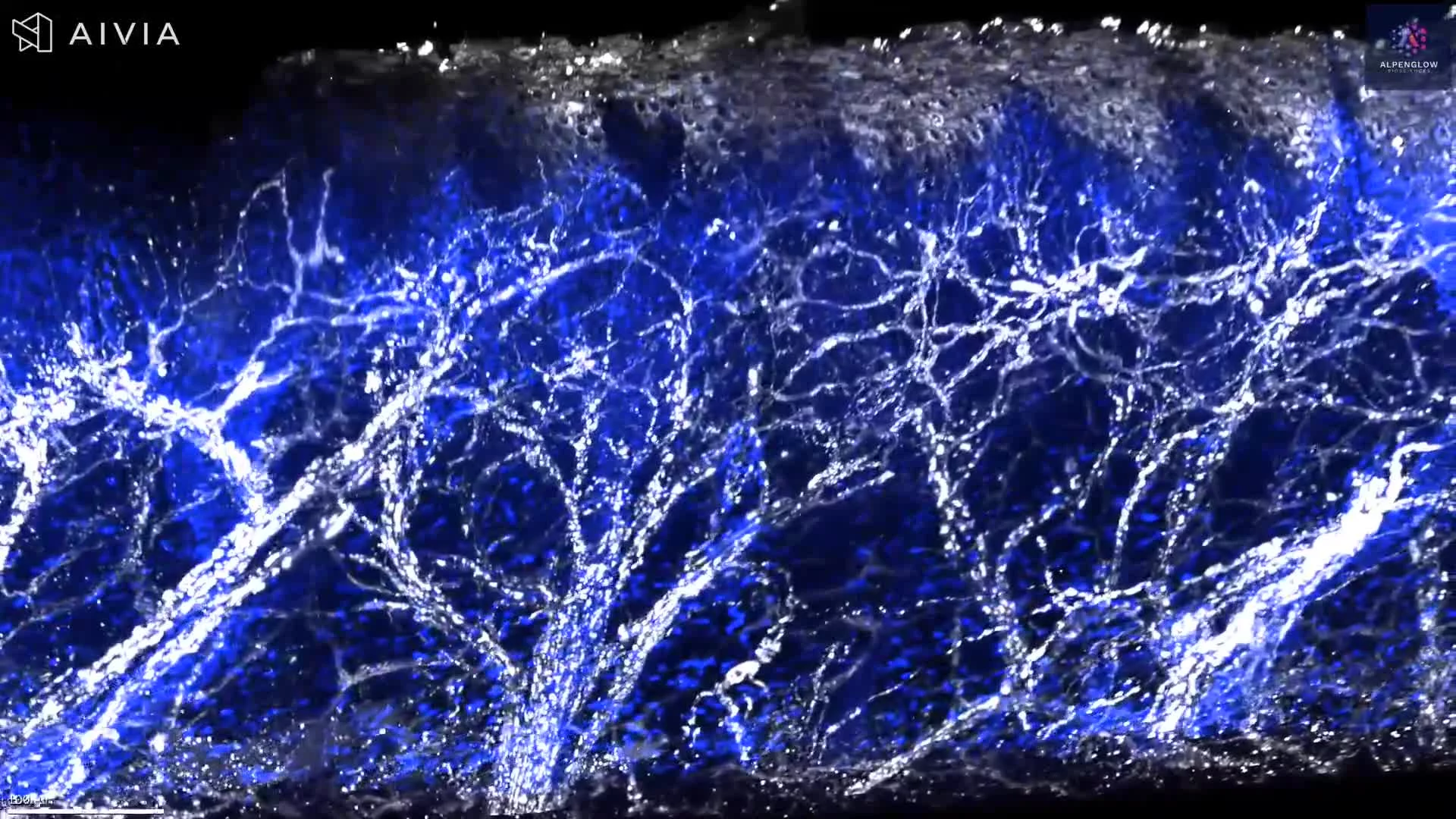
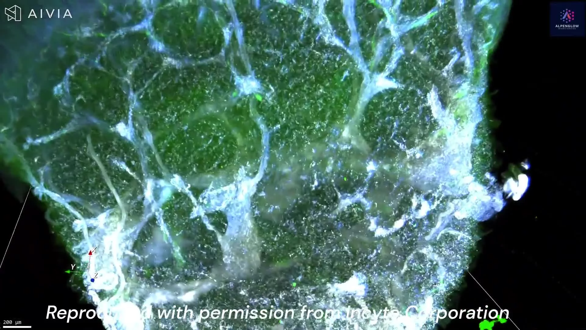
Journey through the delicate nerve structures of the epidermis and dermis at low resolution, and zoom in for an awe-inspiring close-up of lymphocyte distribution around these dynamic networks.
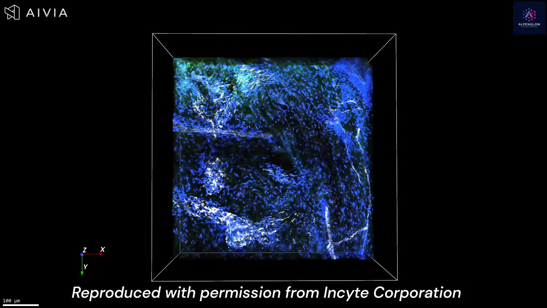
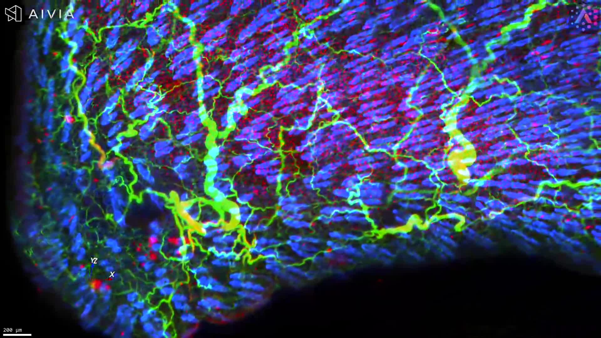
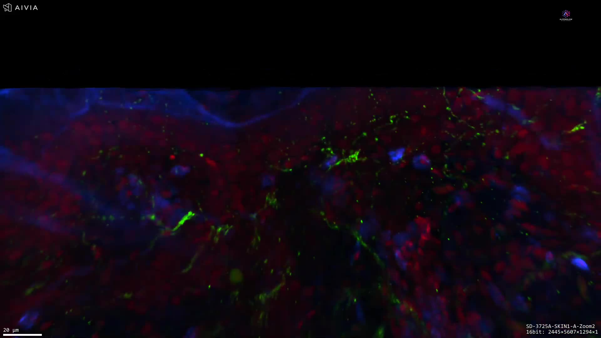
40X image of a lesional Atopic Dermatitis samples stained with PGP9.5 (white) to highlight fine epidermal innervation, TO-PRO-3 (blue) for nuclear detail, and CD3 (green) to illuminate T-Cell infiltration of the Epidermis and Dermis.
Low resolution image of a lesional Atopic Dermatitis sample stained with PGP9.5 (white) to highlight fine epidermal innervation, TO-PRO-3 (blue) for nuclear detail, and CD3 (green) to illuminate T-Cell infiltration of the Epidermis and Dermis.