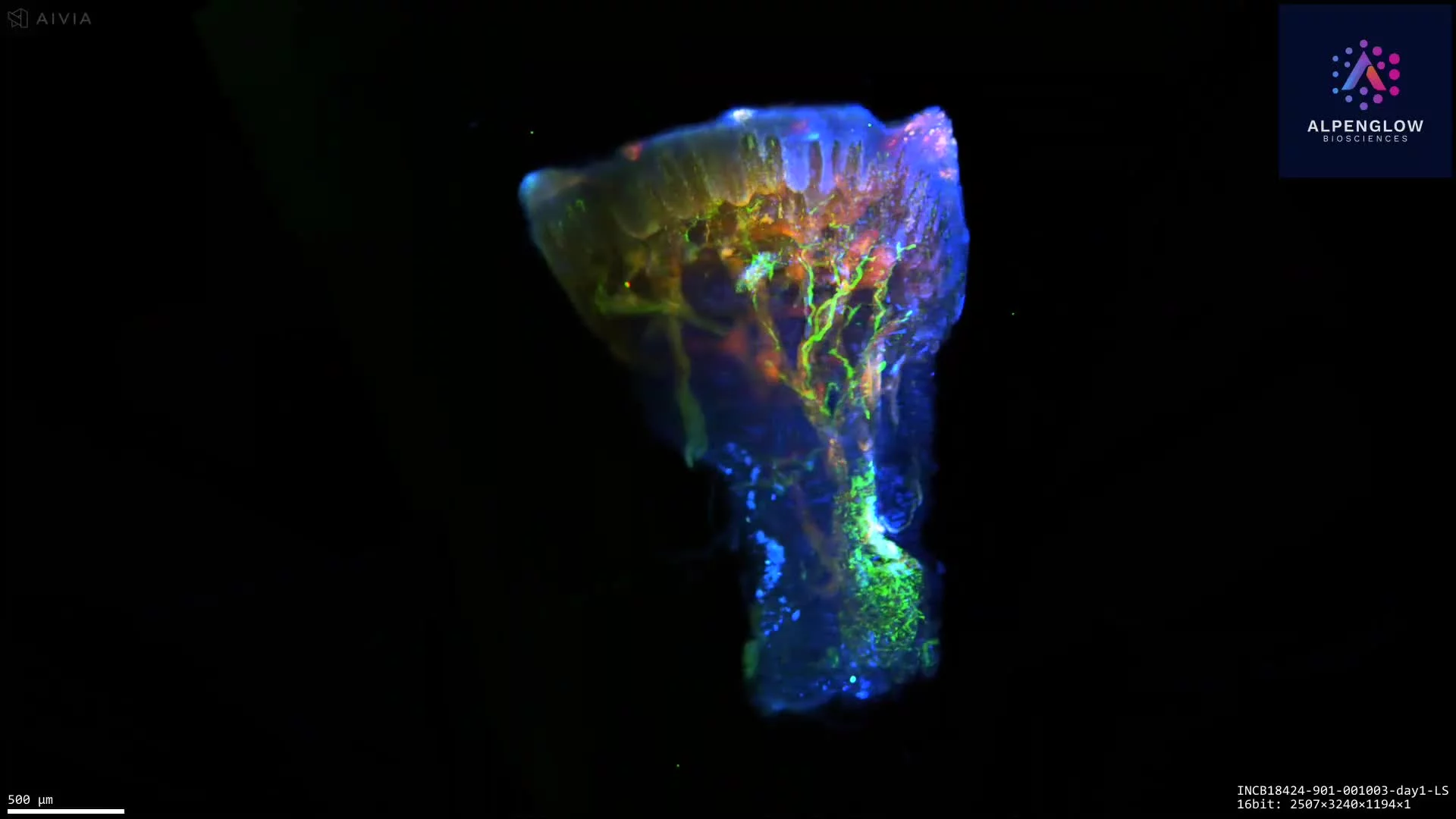
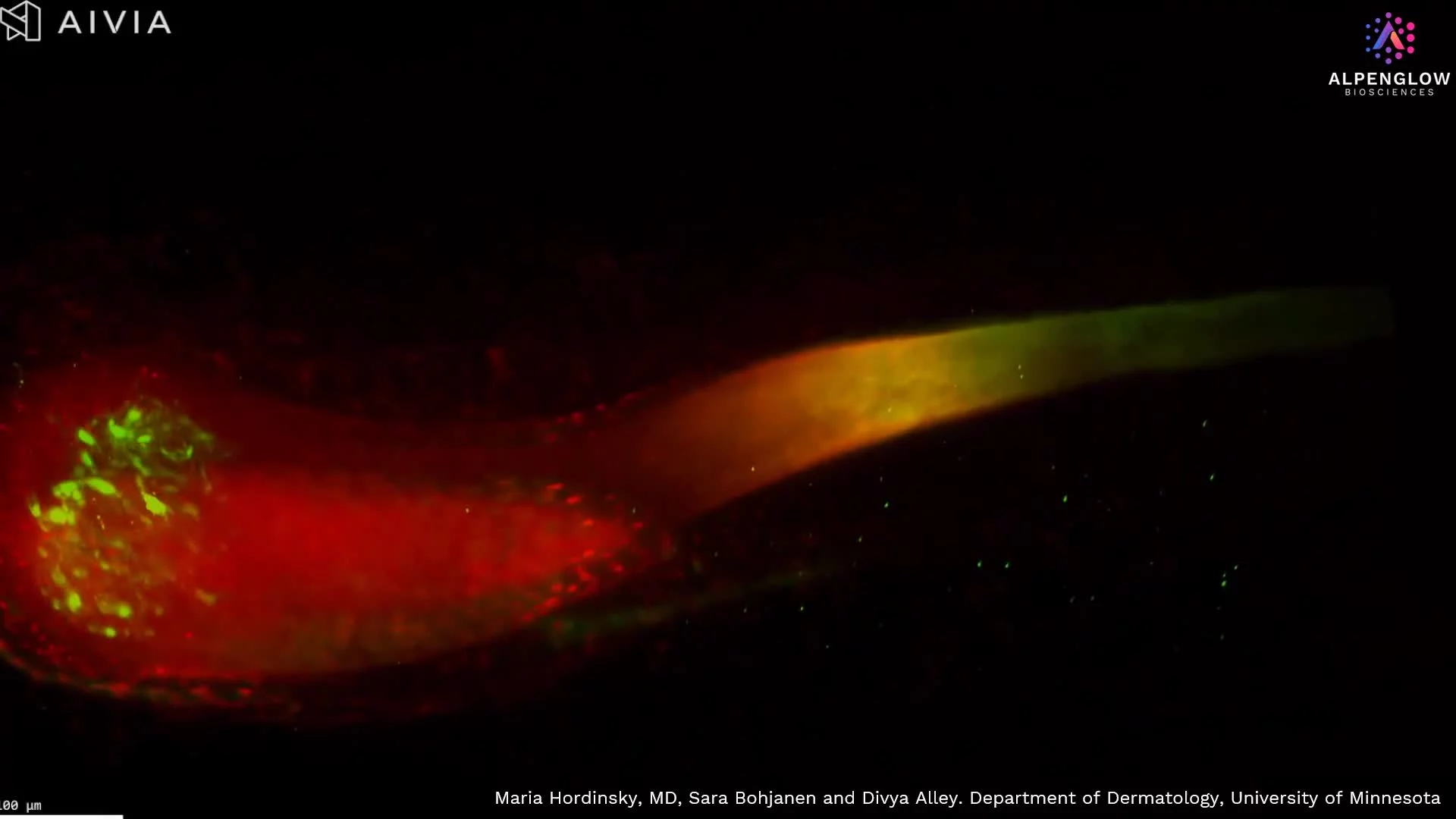
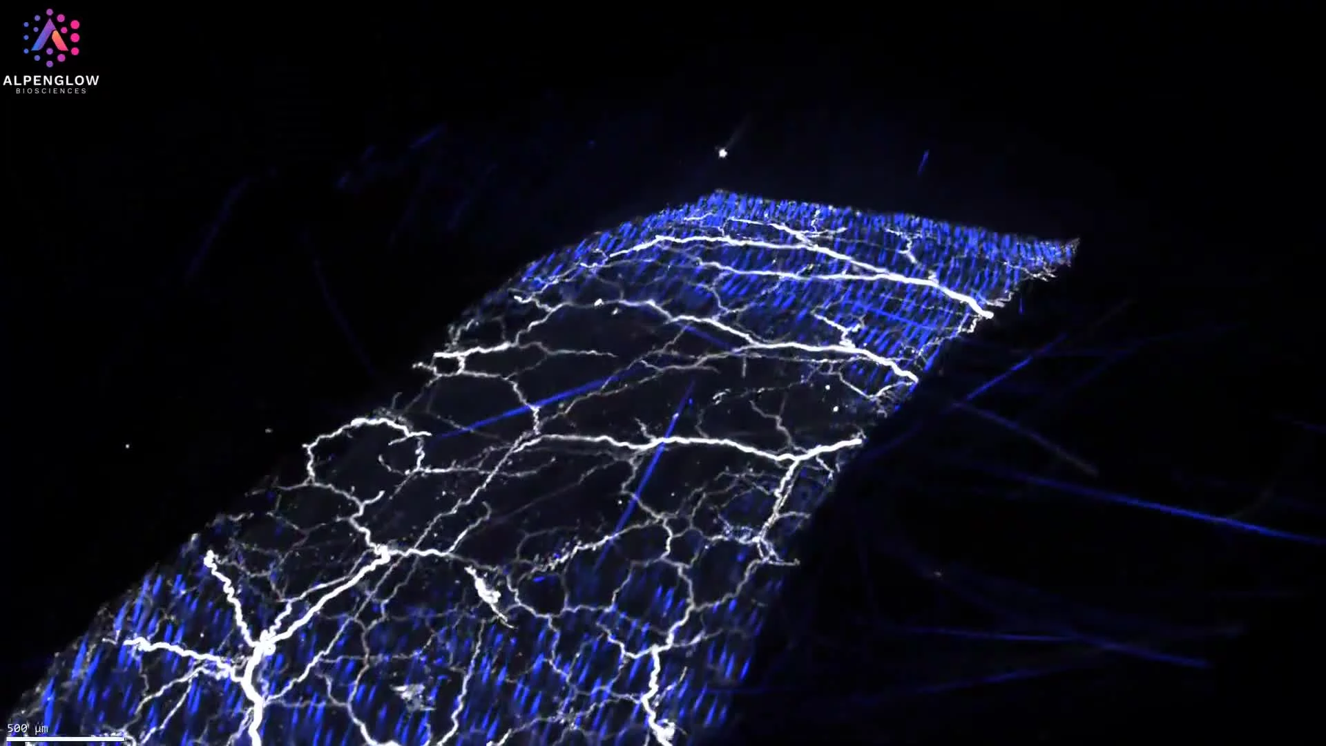
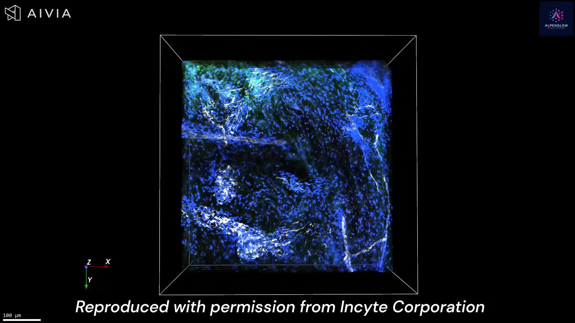
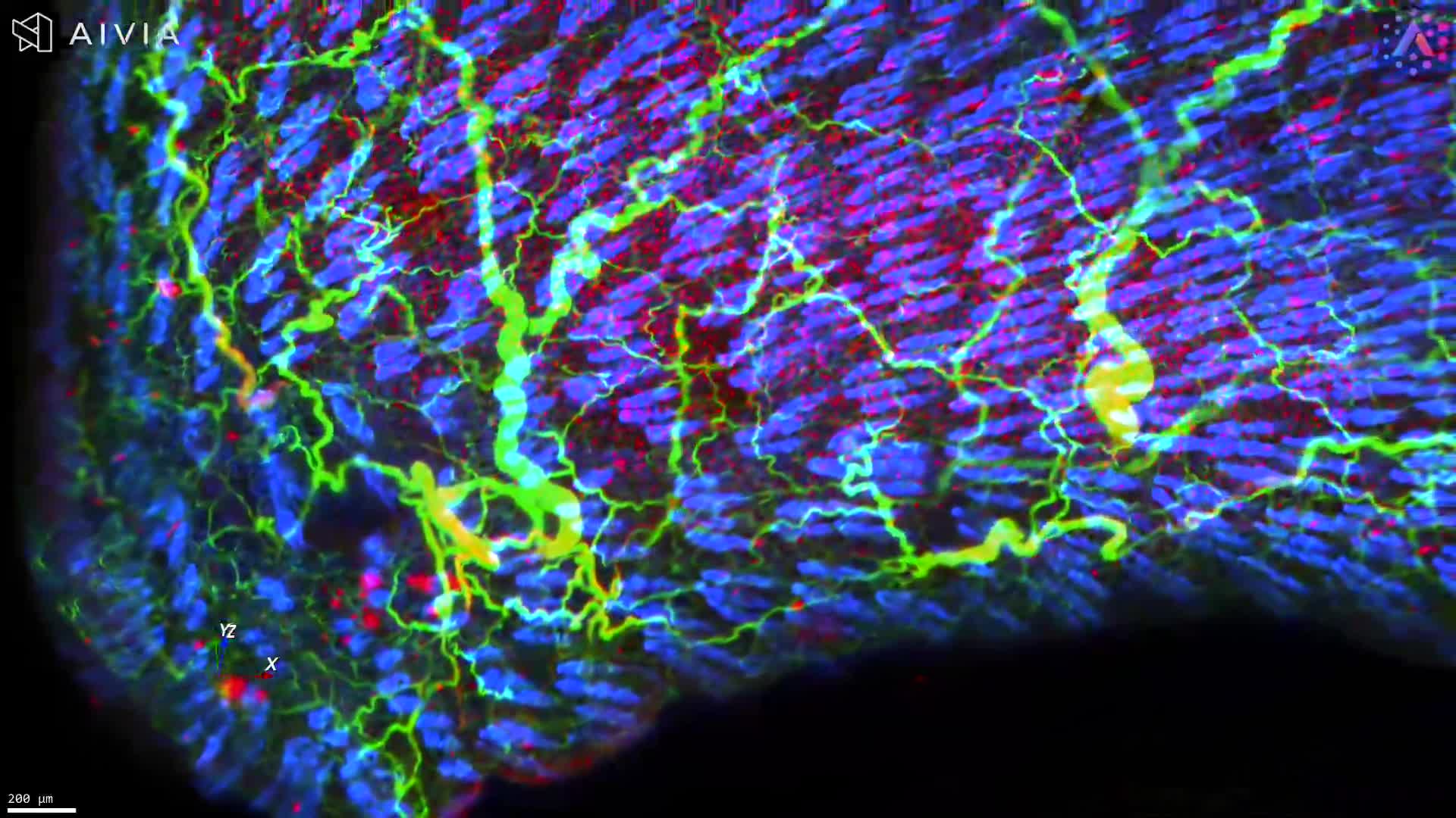
Unlocking the Secrets of Hair Follicles with 3D Imaging
For the first time, a hair follicle can be visualized in its true three-dimensional architecture, from the bulb where growth begins to the dermal papilla, a key signaling center for follicle development. This high-resolution dataset, captured with the Aurora platform and HOTLS microscopy, also reveals the hair plexus, a dense network of sensory nerve fibers critical for touch sensation.
Stains used:
PGP9.5 (Green): Sensory nerve fibers surrounding the follicle
TO-PRO-3 (Red): Cell nuclei distribution
Through advanced 3D fluorescence imaging, combined with data management and AI-powered segmentation, subtle structural changes in the follicle can be detected earlier than with conventional 2D histology. This capability is transforming alopecia research, providing a new window into hair biology, disease mechanisms, and treatment response.
By integrating quantifiable 3D tissue imaging with digital pathology and spatial profiling, researchers gain unprecedented insights into hair follicle biology and inflammatory skin conditions. This breakthrough demonstrates how better imaging leads directly to better insights, and ultimately better treatments.