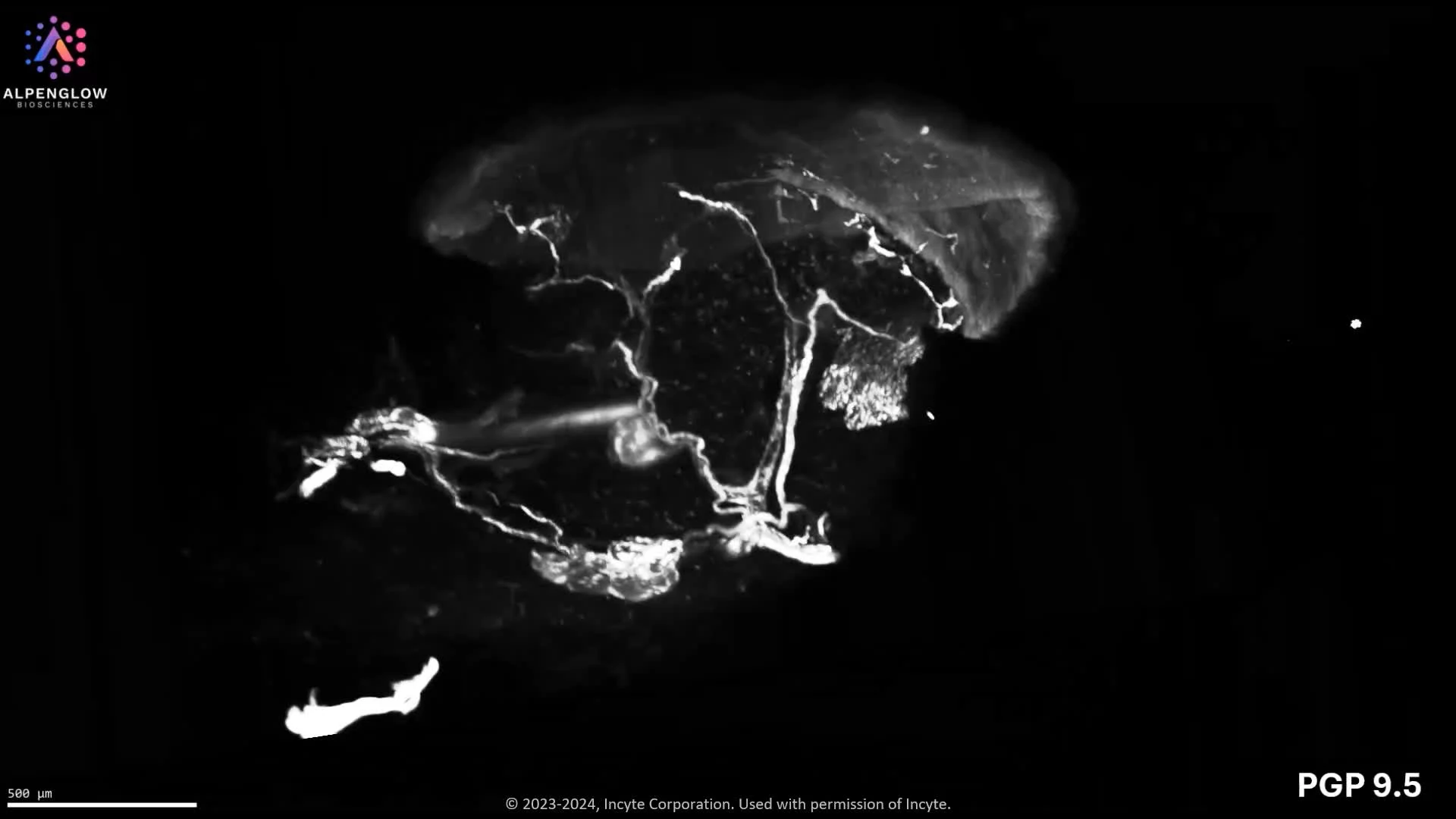
Expanded 3D imaging of mouse kidney. Glomerular and extracellular matrix architecture at subcellular scale
This video showcases three-dimensional imaging of mouse kidney tissue using expansion microscopy, enabling nanoscale visualization of renal structures within an intact volumetric context.
Mouse kidney tissue is physically expanded to an effective refractive index of n = 1.33, allowing optical access to fine structural detail beyond the limits of conventional fluorescence microscopy. Expansion microscopy preserves relative spatial relationships while increasing effective resolution across the entire volume.
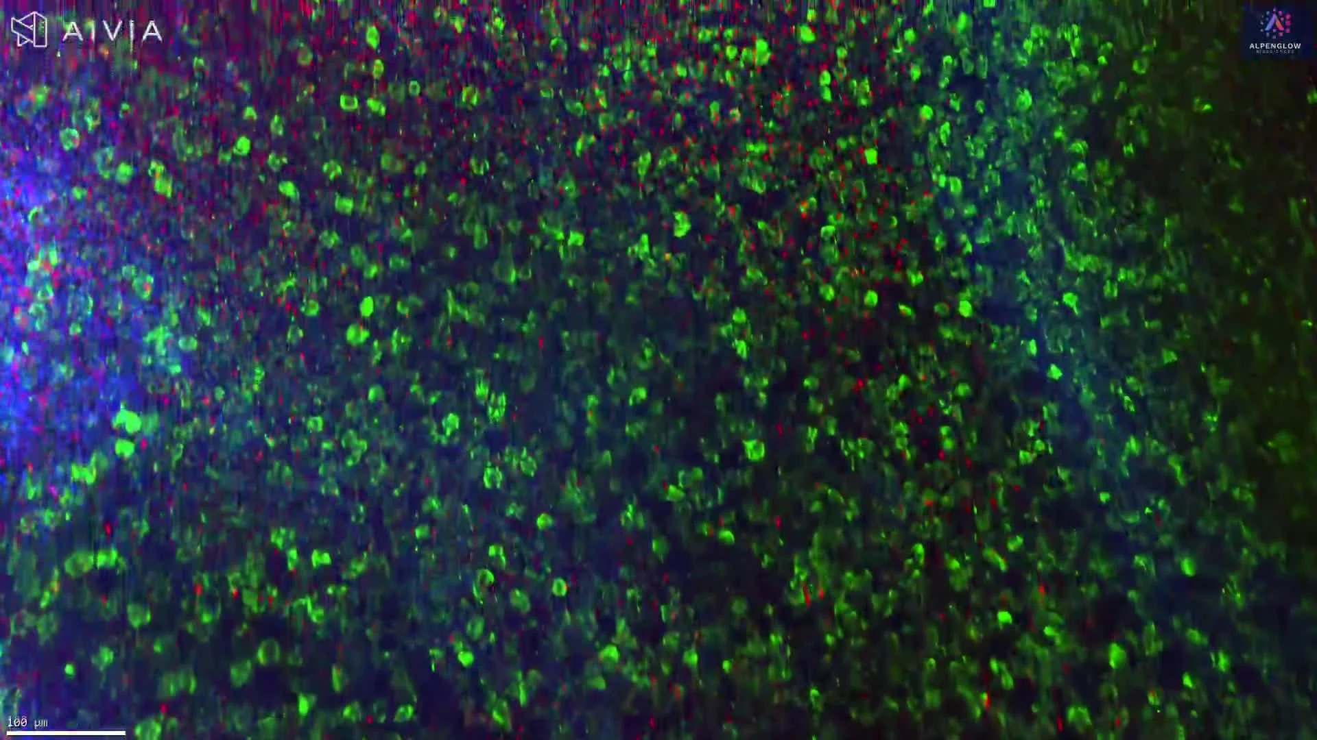
In this example, multiple molecular markers are visualized simultaneously to resolve key kidney compartments.
Nuclei are shown in white.
Collagen IV, a major component of the basement membrane, is shown in blue.
WGA lectin highlights glycoprotein-rich structures in magenta.
Podocalyxin (Podxl) labels glomeruli in green.
Traversing the expanded volume reveals the three-dimensional organization of glomeruli, basement membranes, and surrounding tissue architecture. These spatial relationships are difficult to interpret from thin sections, where structures appear fragmented and disconnected.
This dataset demonstrates how 3D expansion microscopy enables detailed analysis of kidney microanatomy while preserving volumetric context, supporting studies of renal structure, function, and disease mechanisms.
This work was performed in collaboration with Joshua Vaughan and the University of Washington Department of Chemistry.