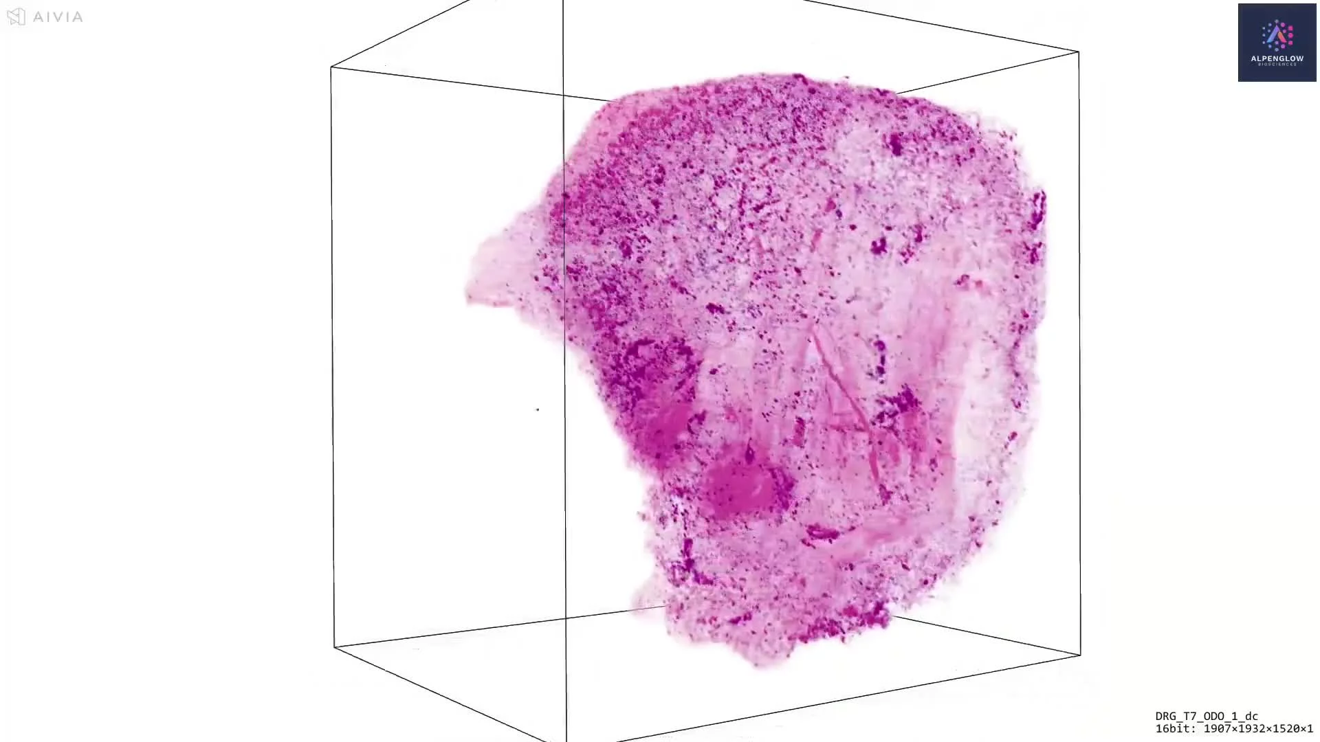
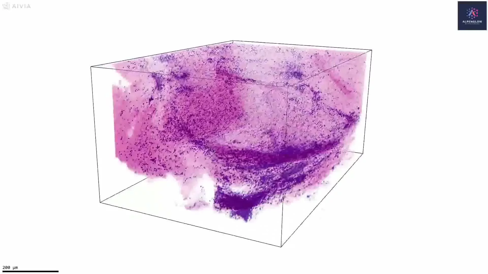
3D Imaging of Human Normal Adjacent Colorectal Cancer
This dataset demonstrates advanced 3D imaging of Human Normal Adjacent Colorectal Cancer tissue, highlighting how whole-section volumetric analysis enables insights far beyond conventional histology.
Workflow:
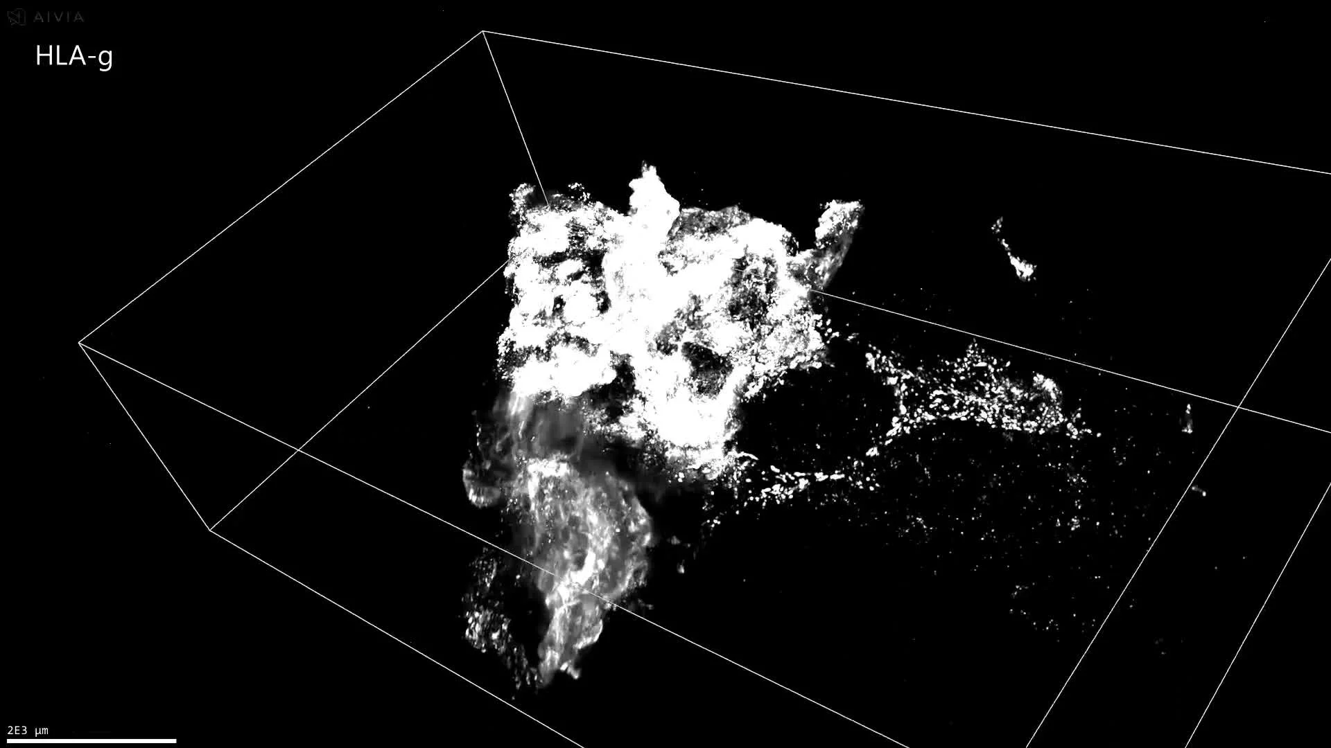
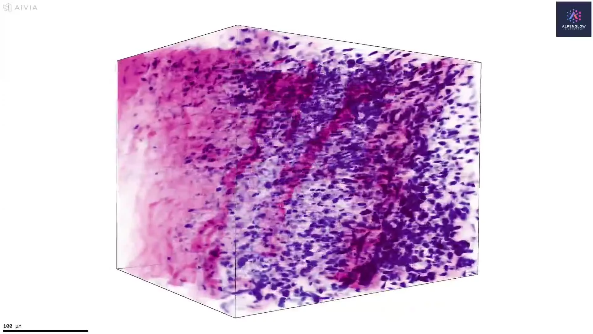
Tissue Preparation: FFPE tissue was deparaffinized, processed, and stained with YO-PRO-1 (blue) for nuclei and anti-CD8 antibody (yellow) for cytotoxic T cells.
Optical Clearing: Modified iDISCO+ protocol ensured full antibody penetration and uniform sample transparency.
Imaging: The sample was imaged at 2 μm/pixel resolution using the Aurora™ 3Di Hybrid Open-Top Light Sheet (HOTLS) microscope.
Analysis: CD8+ lymphocytes were quantified across the entire section (≈6 mm × 4.3 mm × 4.2 mm) with 3Dm data management and 3Dai AI-powered segmentation, enabling precise spatial profiling of immune infiltration.
This workflow provides ground truth 3D histology and quantitative immune mapping, demonstrating the power of digital pathology and 3D spatial biology for oncology. Applications include immune infiltration analysis, biomarker development, and translational research in cancer.