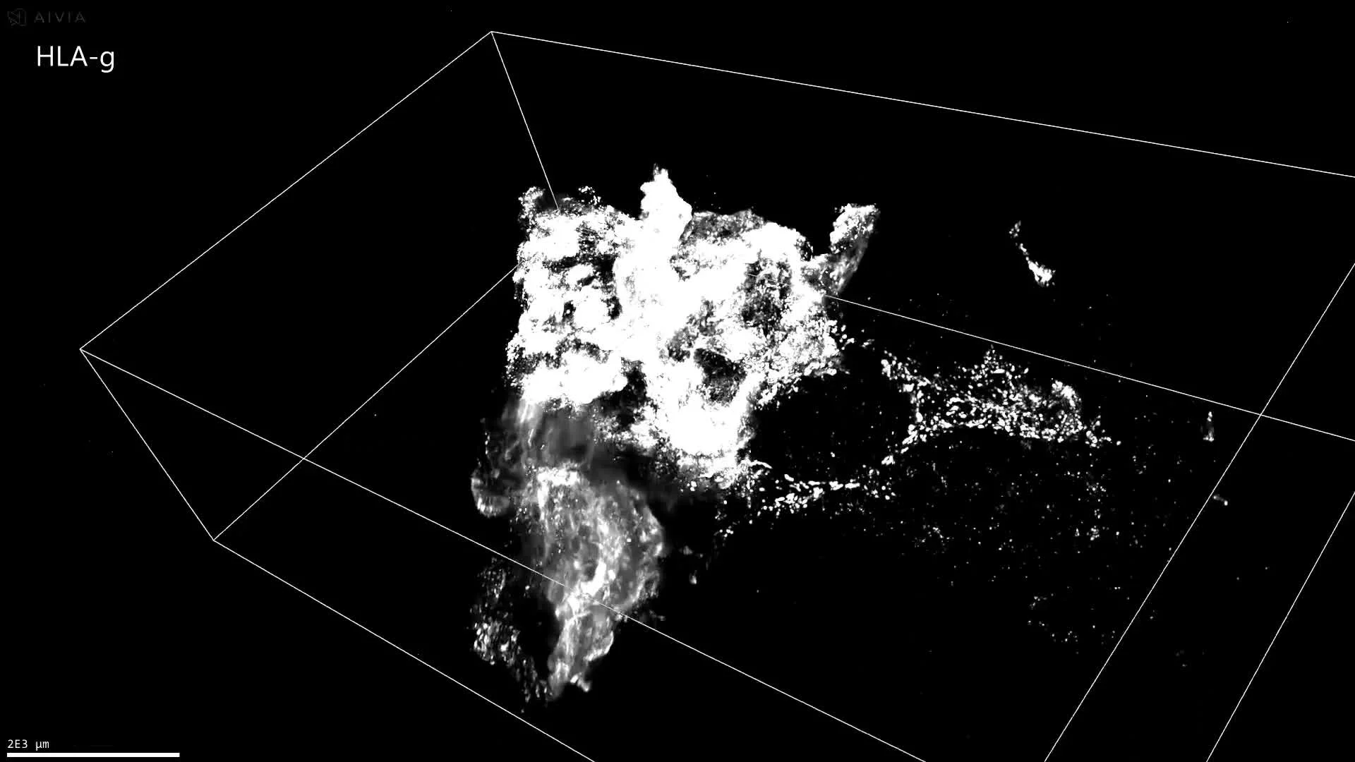
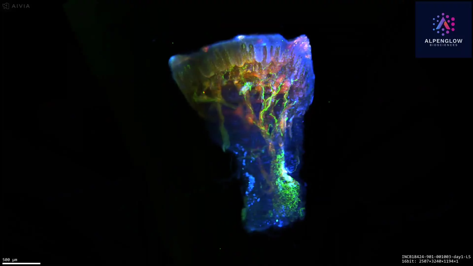
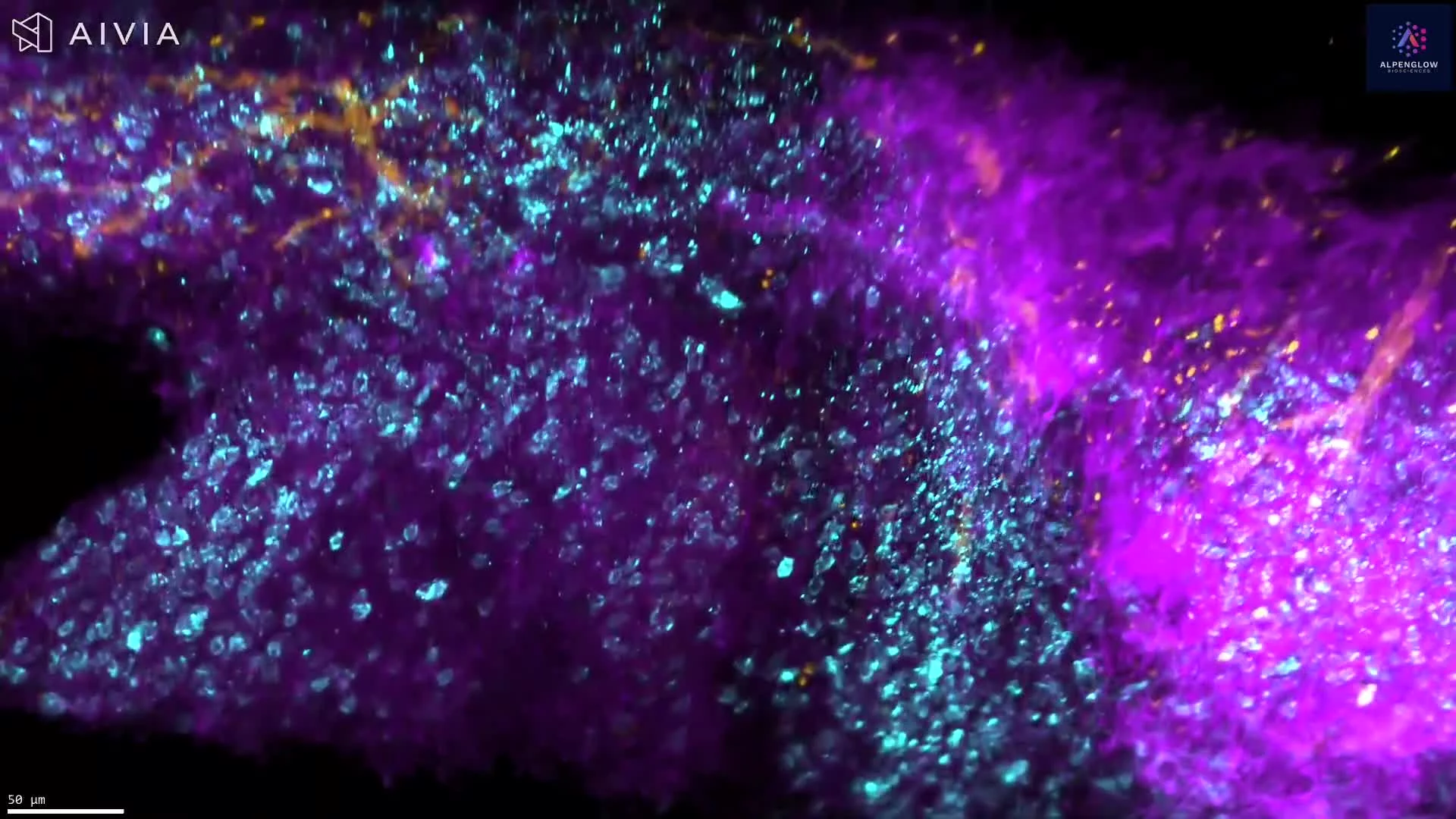
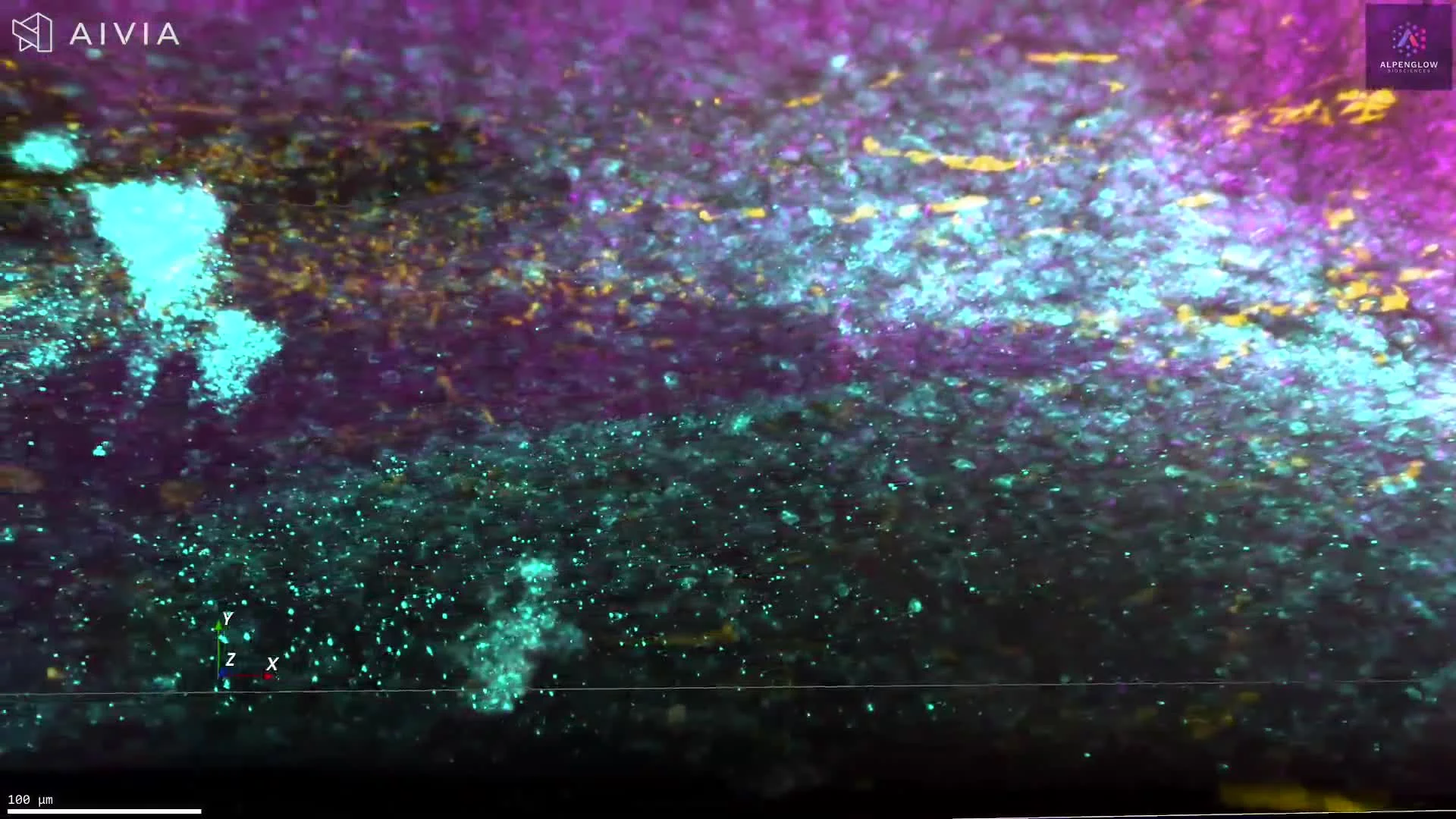
3D Immune Organization in an Intact Mouse Colorectal Tumor
This video presents high-resolution 3D imaging of an intact mouse colorectal tumor, preserving immune organization across the full tissue volume.
B220, shown in green, highlights B-cell-rich regions. CD3, shown in red, identifies T-cell populations, while nuclei are shown in blue to provide cellular and tissue context. Together, these markers reveal how immune cells are distributed, clustered, and spatially organized within the tumor.
The volumetric dataset preserves regional heterogeneity and relationships between B-cell-rich and T-cell-rich areas across depth. With appropriate segmentation, it can support quantitative analysis of immune-cell density, clustering, spatial distribution, and distances between immune populations and surrounding tumor regions.
Explore how 3D tissue imaging supports immuno-oncology research across tumor architecture, immune-cell organization, and spatial relationships.
The tissue was imaged using the Aurora 3D™ Spatial Biology Solution, including the 3Di™ Hybrid Open-Top Light-Sheet microscope.