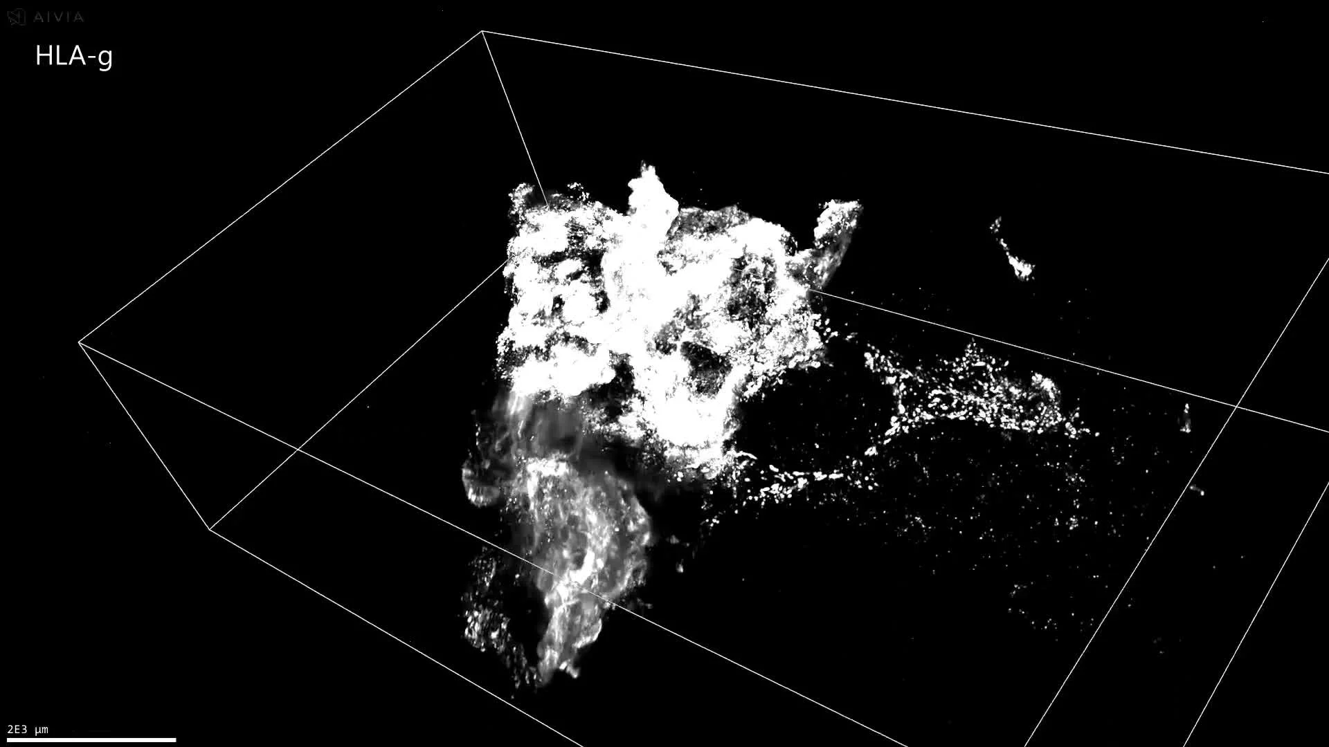
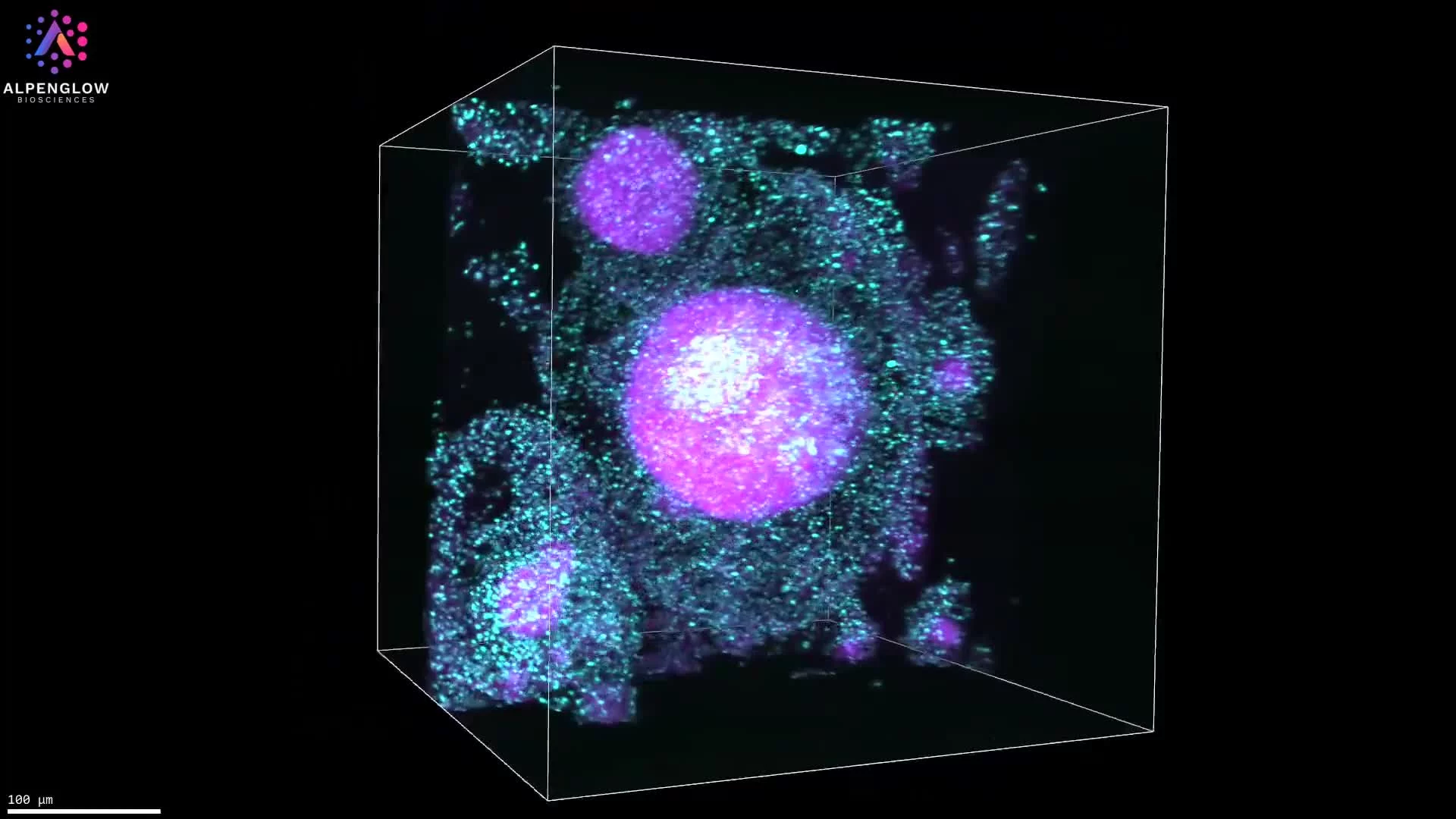
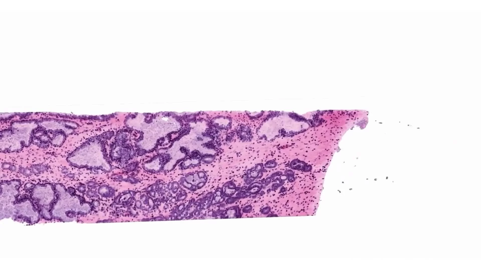
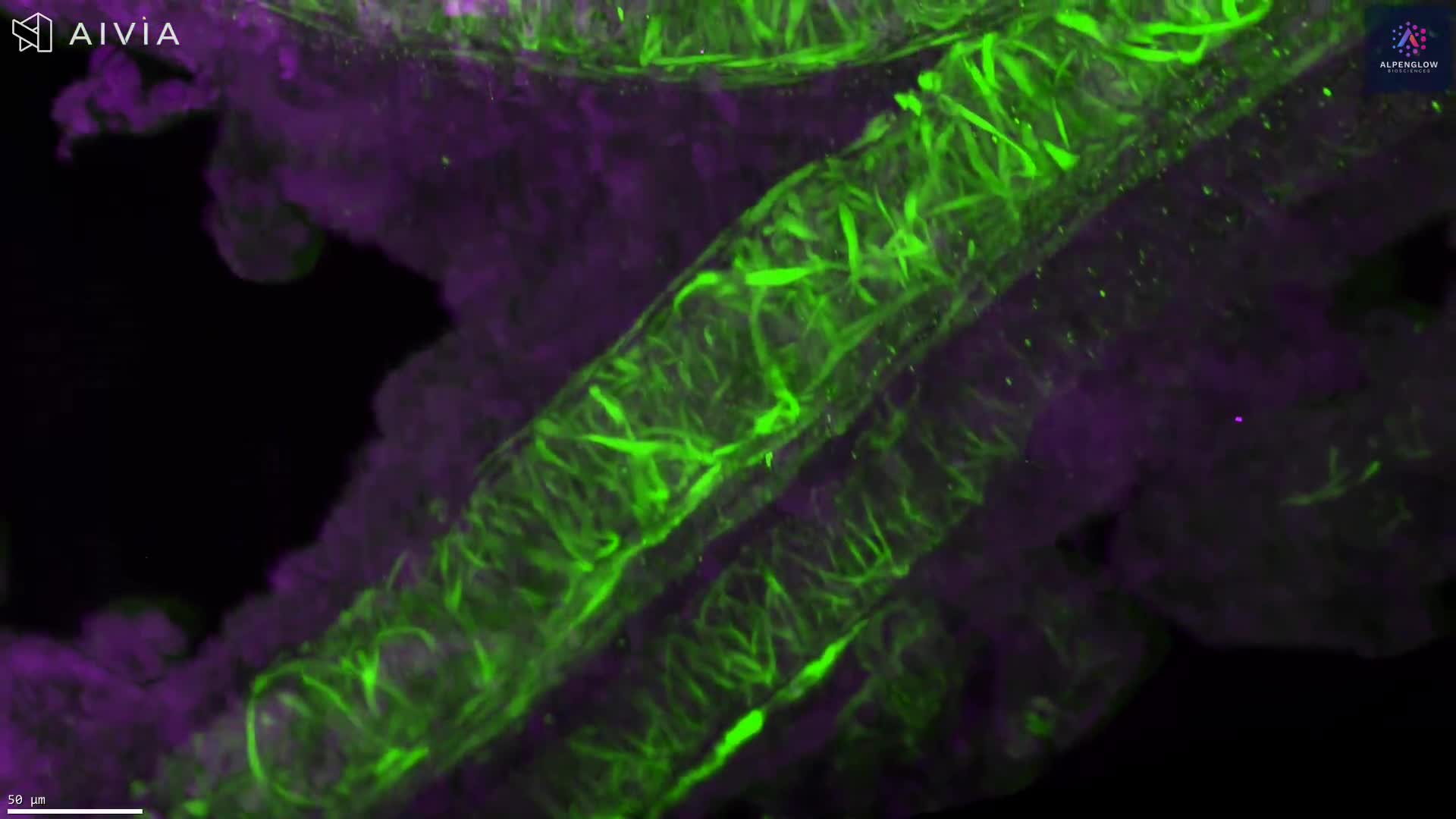
Collagen III Staining and 3D Imaging of Liver Fibrosis
This dataset demonstrates Collagen III staining of a human liver sample after tissue clearing and high-resolution 3D imaging on the Aurora™ 3Di Hybrid Open Top Light Sheet (HOTLS) microscope. By combining fluorescence labeling with volumetric imaging, the dataset preserves intact tissue structure while highlighting fibrotic regions across the liver sample.
Following imaging, advanced image analysis workflows and a pixel classifier were applied to segment fibrotic tissue. This approach highlights fibrosis in its full three-dimensional context, providing quantitative data that far surpasses traditional 2D histology.
Integration with 3Dm data management and 3Dai AI-powered segmentation enables reproducible classification and quantification of fibrosis across the entire liver volume. These insights are invaluable for digital pathology, spatial profiling, and translational research in hepatology, supporting studies of liver fibrosis, steatosis, and related metabolic and inflammatory conditions.
Contact us to learn how Alpenglow’s 3D spatial biology platform can advance your research with large-scale, quantitative imaging datasets.