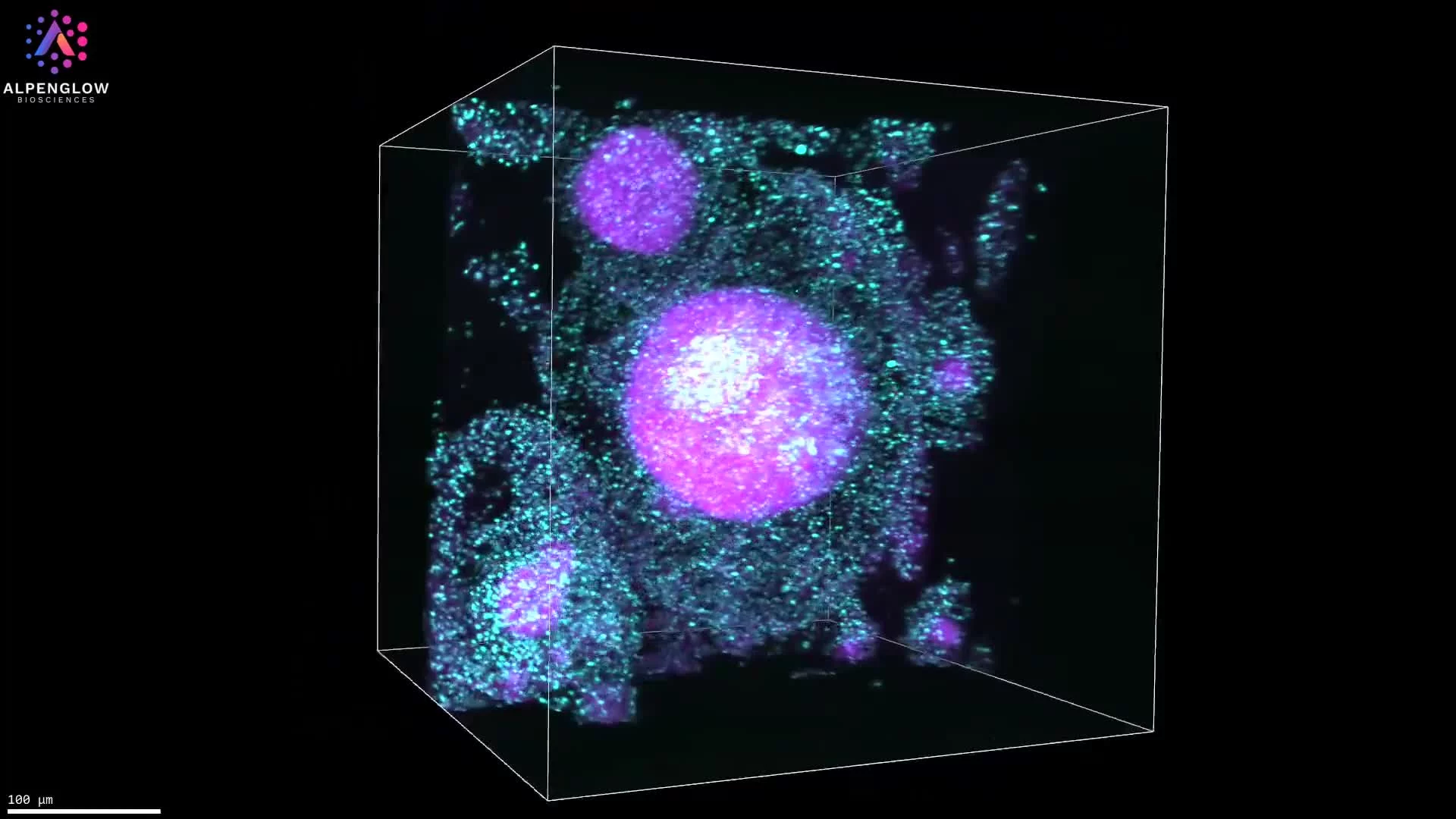
Exploring Prostate Organoids in 3D with LUMI
Prostate organoids are complex 3D models that demand a high-resolution, volumetric approach to imaging. This video demonstrates how LUMI (Light-Sheet User Microscope Interface) simplifies the process by combining low-resolution scanning with smart region selection and ultra-high-resolution imaging of specific areas.
Workflow with LUMI Smart Microscopy:
Fast pre-scan: Capture the entire organoid sample at low resolution.
ROI definition: Select regions of interest directly on the whole 3D raw image data.
Multi-ROI scanning: Efficiently image multiple organoids using LUMI’s multi-well setup.
In this example, prostate organoids are stained with TO-PRO-3 and eosin, then pseudocolored to enhance structural details. The result is a precise, multi-scale analysis of organoid organization, bridging low-resolution overview with single-cell clarity.
By pairing LUMI’s interface with the Aurora™ 3Di Hybrid Open Top Light Sheet (HOTLS) microscope, researchers gain seamless control over imaging workflows. Integration with 3Dm data management and 3Dai AI-powered segmentation further enables quantitative analysis of organoid growth, structure, and cellular interactions.
Applications extend to cancer biology, drug testing, and translational research, where prostate organoids serve as powerful disease models. LUMI accelerates these workflows, making 3D organoid imaging more accessible, reproducible, and insightful.