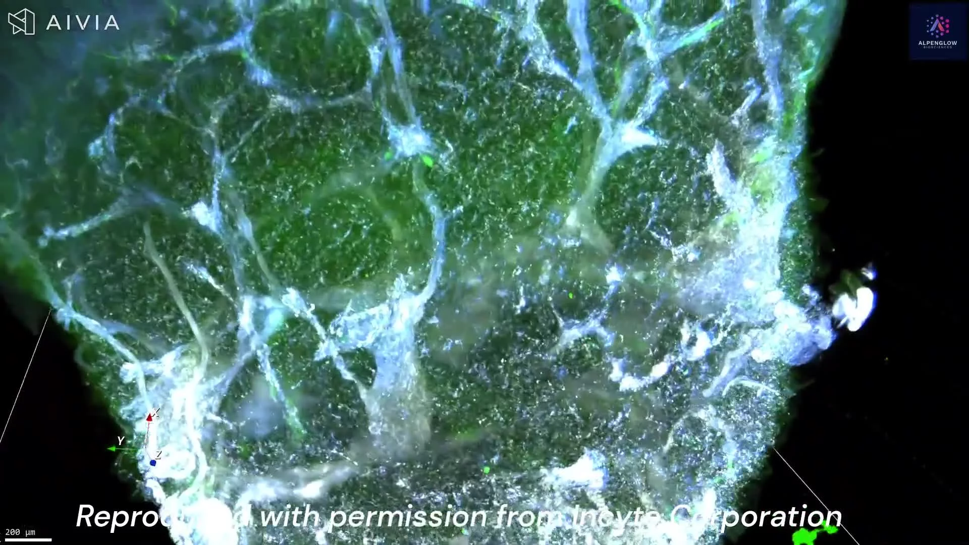
High-Resolution 3D Imaging of a Hair Follicle in Hidradenitis Suppurativa
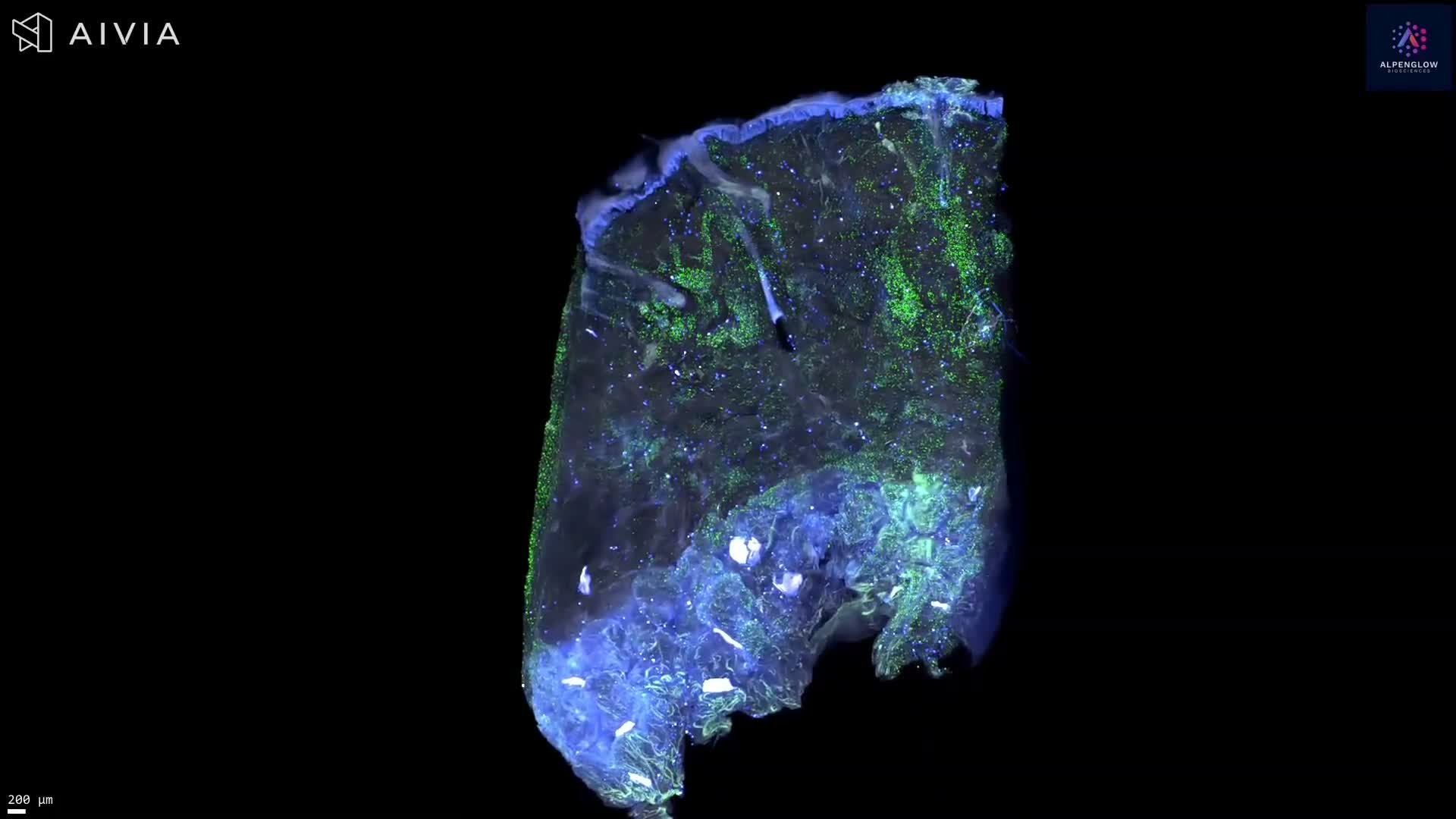
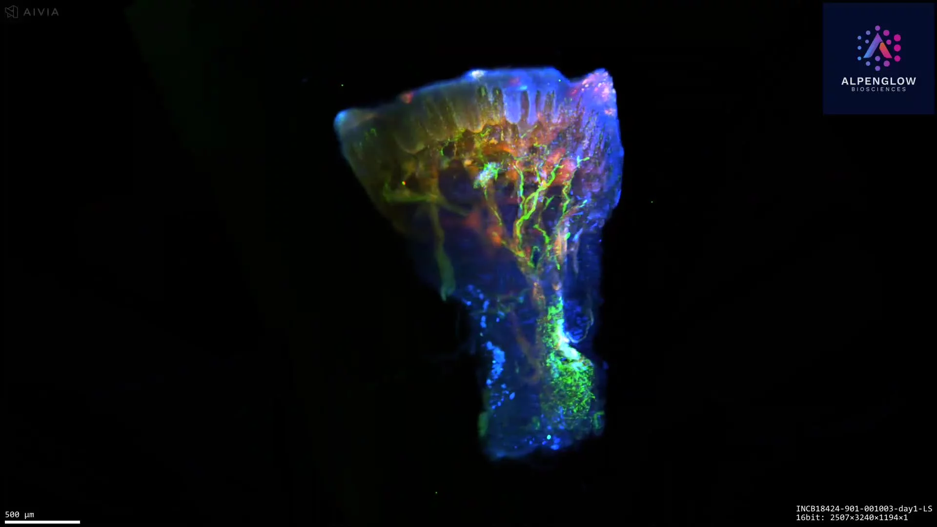
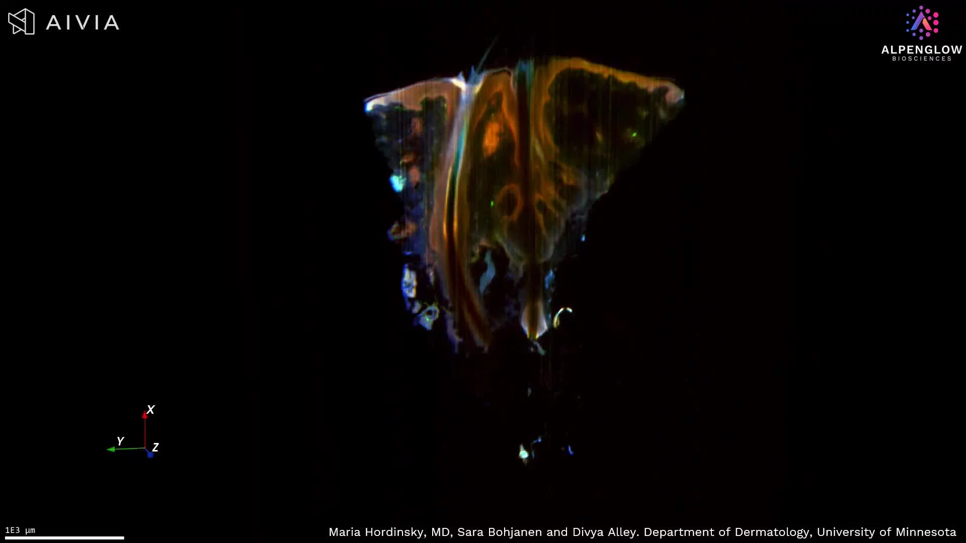
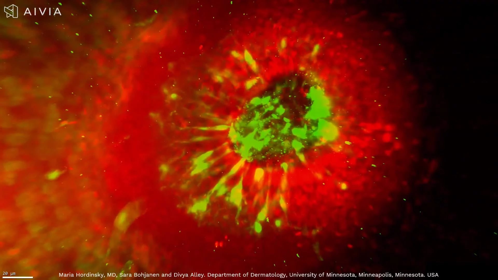
This dataset presents high-resolution 3D fluorescence imaging of a hair follicle within an intact hidradenitis suppurativa (HS) skin biopsy, revealing follicular architecture, surrounding nerve networks, and individual cells throughout the imaged tissue volume.
The visualization begins with a 3D reconstruction of the complete hair follicle, showing its morphology and the intricate distribution of nerve fibers and mast cells in the surrounding tissue. Virtual 2D sectioning enables detailed examination of cellular structures at different depths without physically sectioning or destroying the specimen. The dataset is then visualized again in 3D to reveal the spatial organization of these structures.
Fluorescence markers:
- TO-PRO-3 (blue): Cell nuclei
- PGP9.5 (white): Nerve fibers
- Tryptase (green): Mast cells
The combination of high spatial resolution and volumetric imaging enables quantitative assessment of follicular morphology, cell counts and densities, nerve architecture, and distances between cells and nerve fibers.
This approach supports the investigation of follicular structures and neuroimmune interactions in HS while preserving their spatial relationships within the intact tissue.