3D Visualization of Hair Follicles, Nerves, Mast Cells, and Tunnels in Hidradenitis Suppurativa
Hidradenitis suppurativa (HS) is a chronic inflammatory skin disease characterized by recurrent inflammatory lesions, tissue remodeling, and the formation of epithelialized tunnels. Understanding the three-dimensional organization of these structures and their surrounding microenvironment is important for investigating disease pathology.
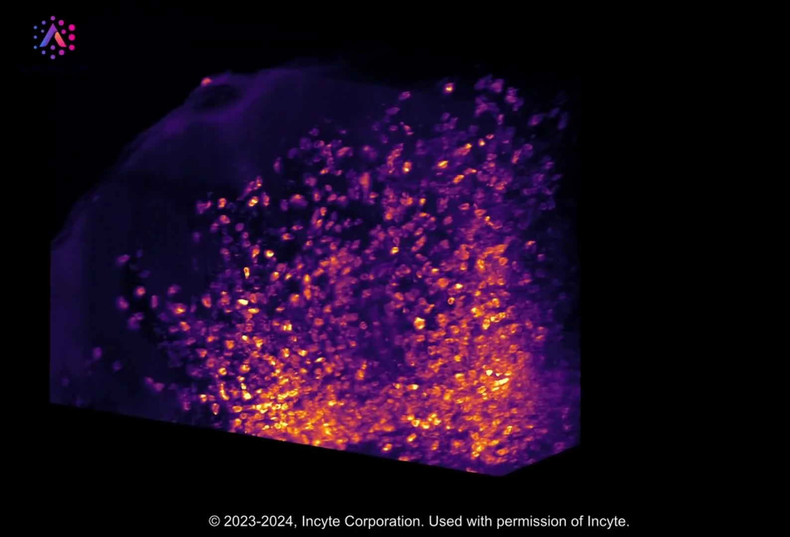
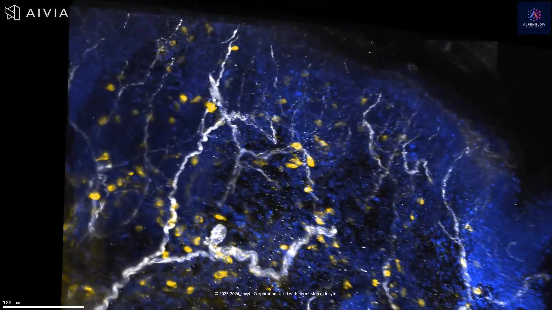
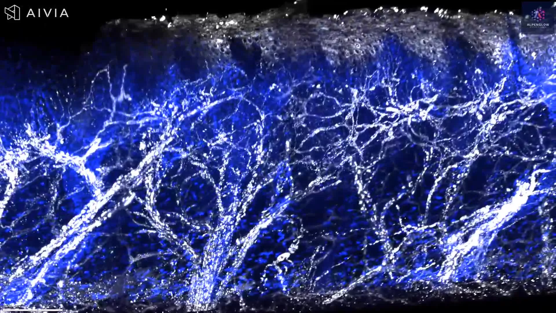
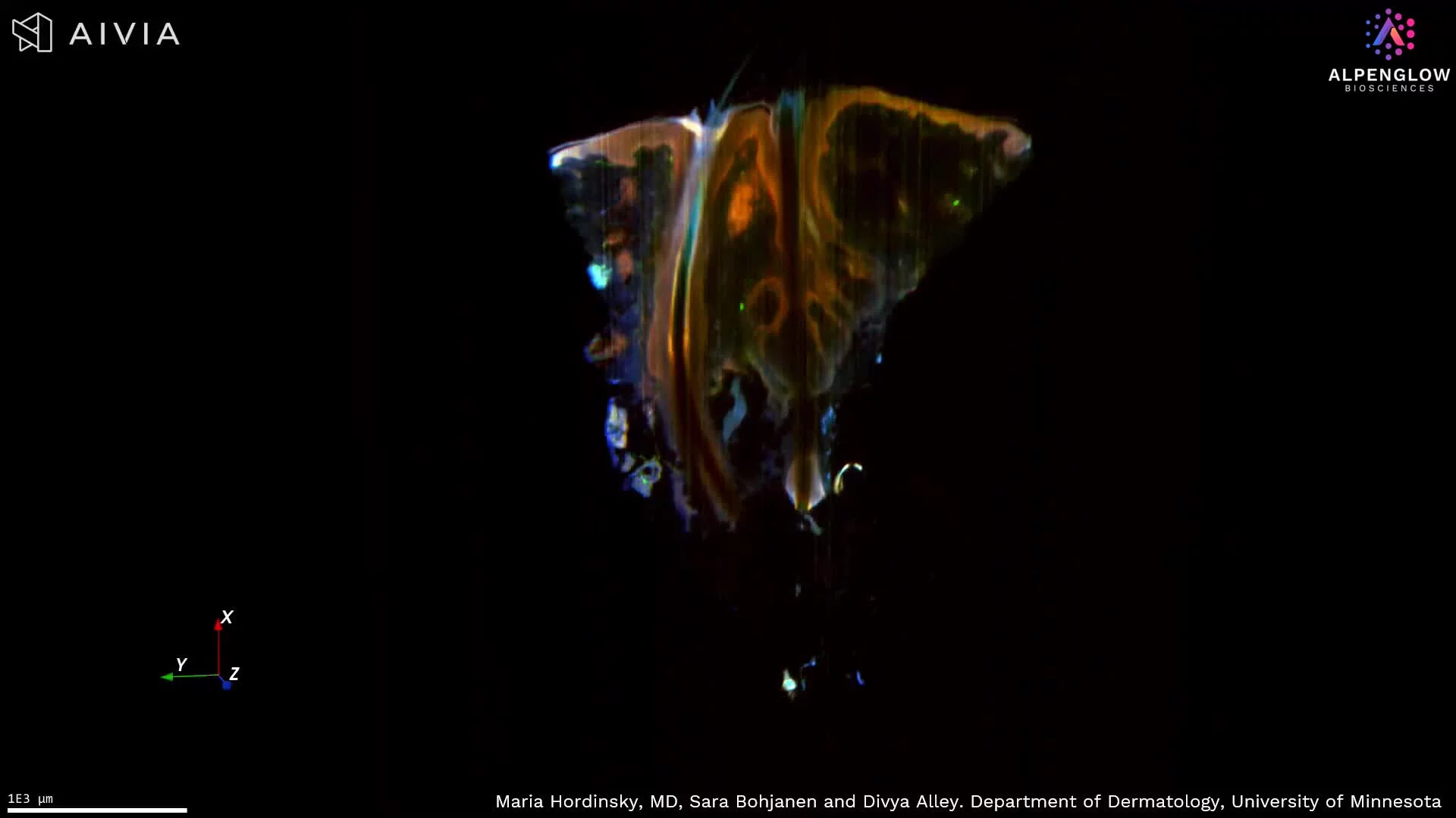
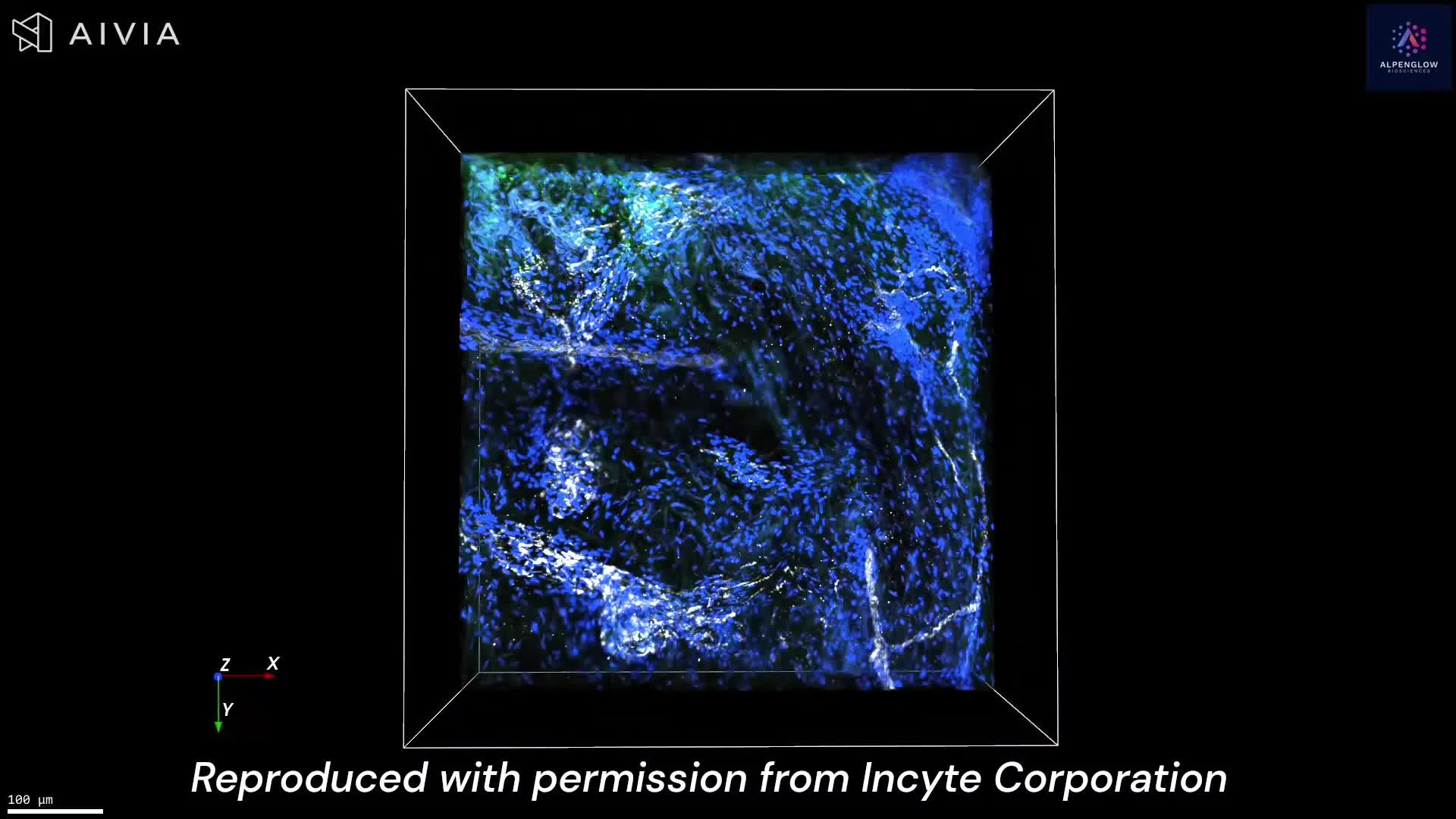
This video presents a volumetric visualization of an HS skin biopsy acquired using 3D light-sheet fluorescence microscopy. Three fluorescent markers highlight distinct tissue components:
- Blue: Cell nuclei
- White: PGP9.5-positive nerve fibers
- Green: Tryptase-positive mast cells
The visualization follows hair follicles through the tissue volume, revealing their architecture and spatial relationships with surrounding nerve fibers and mast cells. In conventional two-dimensional histological sections, follicular structures may be only partially represented, depending on the orientation and position of the section.
As the visualization progresses through the biopsy, an HS tunnel becomes visible. Three-dimensional imaging allows its morphology and continuity to be examined alongside the distribution of nearby follicles, nerves, and mast cells.
By preserving the spatial relationships between these structures, volumetric imaging provides a framework for investigating tissue organization, neuroimmune interactions, and inflammatory remodeling in HS.
Such analyses may contribute to a better understanding of the tissue microenvironment associated with tunnel formation and disease progression.