ESDR 2026 · Heidelberg · 9–12 September
See more of skin biology at ESDR 2026
Join our lunch symposium on 3D dermatopathology, or meet privately with Alpenglow to discuss how whole-tissue imaging and Summit AI™ can transform intact skin biopsies into quantitative evidence for dermatology research and development.
Plan your ESDR visit
Choose how you want to connect
Attend the scientific session for a broader view of 3D dermatopathology, then use a private meeting to explore the tissue questions and study requirements specific to your program.
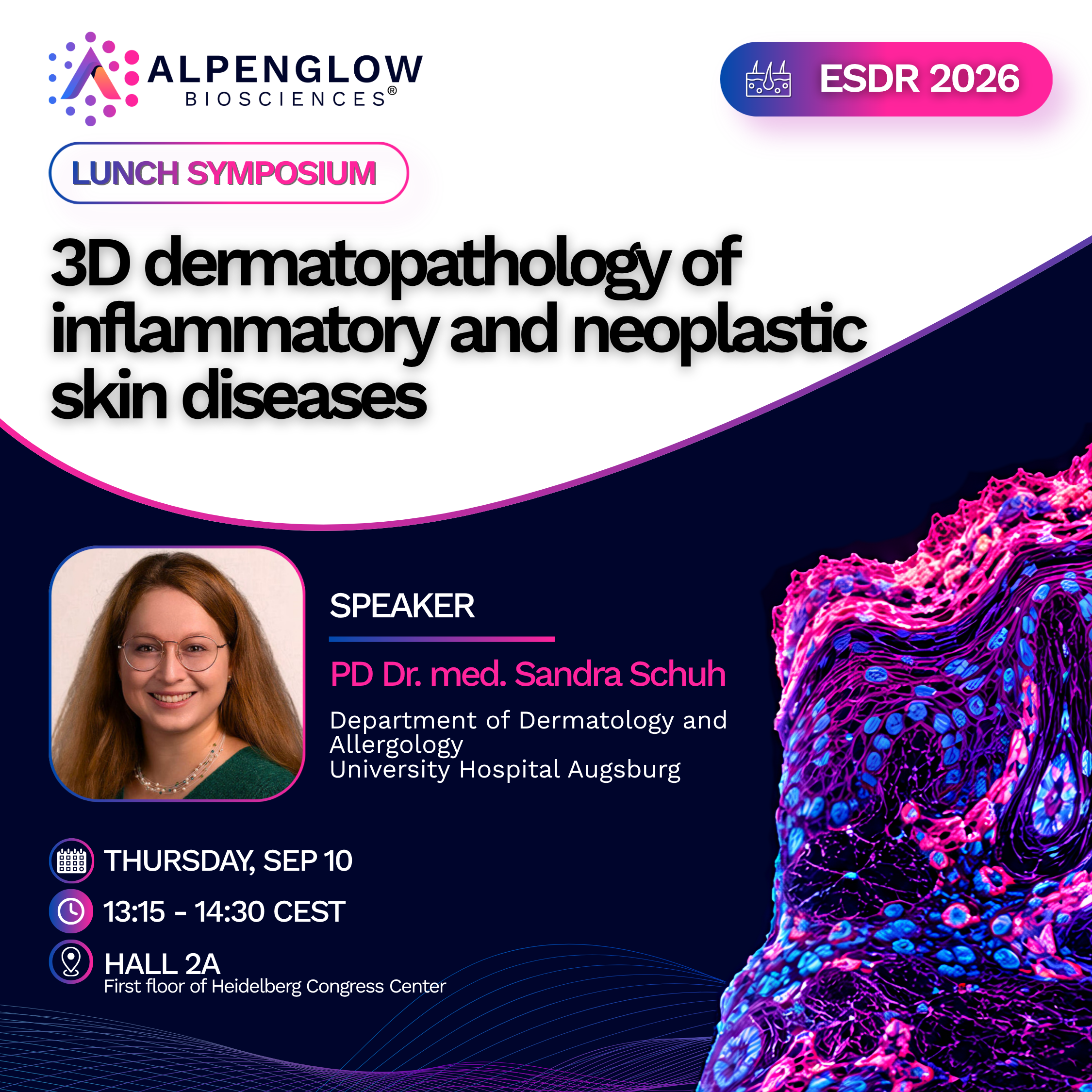
3D dermatopathology of inflammatory and neoplastic skin diseases
Explore how 3D analysis of intact tissue can reveal disease-associated architecture, cellular organization, heterogeneity, and pathological features that may be fragmented or missed across individual tissue sections.
Private discussions
Bring your tissue question to a 1:1 meeting
Meet with the Alpenglow team to discuss how intact-tissue imaging and AI-powered analysis could fit the biological questions, samples, and development decisions behind your study.
- Discuss sample formats, markers, cohorts, and study design
- Define relevant spatial and volumetric measurements
- Explore 3D histology services or an in-house imaging workflow
- Discuss how Summit AI™ can connect tissue data and teams
Select an available time during ESDR through the booking calendar.
Turn complex tissue data into measurable disease biology
Whole-biopsy imaging provides more tissue context, but also creates larger and more complex datasets. Summit AI™ connects full-sample imaging, AI-powered analysis, spatial measurements, and study metadata in one quantitative analysis workflow.
Full-sample analysis
Analyze intact tissue volumes while preserving complete architecture and spatial context.
Quantitative tissue signatures
Convert cells, structures, regions, and relationships into standardized measurements.
Multimodal context
Connect 3D imaging with pathology, molecular data, clinical information, and study metadata.
Cohort-level comparison
Compare measurable tissue patterns across patients, groups, treatments, lesions, and timepoints.
From biopsy to evidence
Move beyond an isolated tissue section
Alpenglow combines whole-tissue 3D imaging with centralized AI-assisted analysis to turn intact skin biopsies into quantitative information for dermatology research and translational programs.
Preserve whole-biopsy context
Follow inflammatory patterns, follicles, vessels, nerves, lesions, tissue remodeling, and connected structures throughout the tissue volume.
Measure disease-associated biology
Quantify cell density, structural volume, spatial relationships, clustering, regional variation, and patient-level heterogeneity.
Connect results across teams
Generate standardized outputs that support pathology, biomarker, translational, discovery, and data teams working from the same tissue evidence.
Choose the right access model
Use Alpenglow as an end-to-end 3D histology service, bring the imaging workflow into your facility, or combine imaging with Summit AI™ for full-sample quantitative analysis.
Meet us in Heidelberg
Make 3D tissue analysis part of your ESDR agenda
Join the lunch symposium for the scientific perspective, or reserve a private conversation focused on your samples, program, and quantitative tissue-analysis goals.